399
Views
0
CrossRef citations to date
0
Altmetric
Research Article

Triflavin, an αIIβ3 disintegrin, inhibits cell adhesion and protein kinase C-α translocation in vascular smooth muscle cells

, , , &
Pages 948-954 | Received 07 Apr 2008, Accepted 30 May 2008, Published online: 07 Aug 2009

Figures & data

Figure 1. Morphological photograph of primary cultured rat aortic vascular smooth muscle cells. The black bar represents 100 μm.
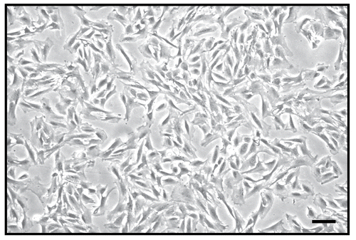
Figure 1.  Morphological photograph of primary cultured rat aortic vascular smooth muscle cells. The black bar represents 100 μm.

Figure 2. Microscopic analysis of the inhibitory effect of triflavin in vascular smooth muscle cell (VSMC) adhesion to immobilized fibronectin. Cells (105 cell/well) were pretreated with (A) PBS or triflavin (B, 10; C, 20; D, 50 μg/mL) and incubated for 1 h to allow adhesion to fibronectin (50 μg/mL)-precoated wells. Non-adherent cells were rinsed off by PBS, and the remaining cells were fixed and photographed under a phase-contrast microscope. The black bar represents 50 μm.
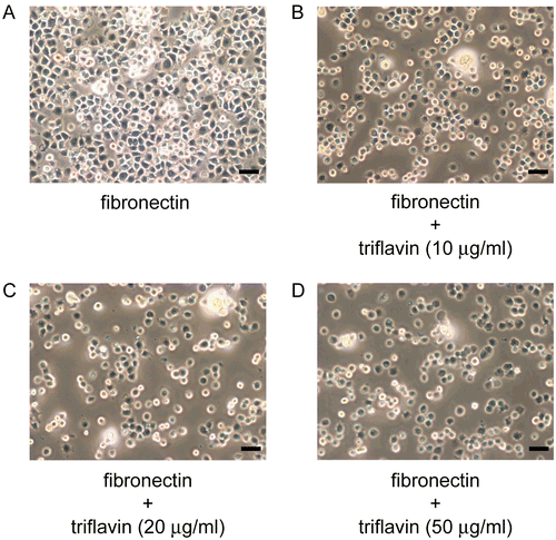
Figure 2.  Microscopic analysis of the inhibitory effect of triflavin in vascular smooth muscle cell (VSMC) adhesion to immobilized fibronectin. Cells (105 cell/well) were pretreated with (A) PBS or triflavin (B, 10; C, 20; D, 50 μg/mL) and incubated for 1 h to allow adhesion to fibronectin (50 μg/mL)-precoated wells. Non-adherent cells were rinsed off by PBS, and the remaining cells were fixed and photographed under a phase-contrast microscope. The black bar represents 50 μm.

Figure 3. Effects of triflavin on vascular smooth muscle cell (VSMC) adhesion to immobilized fibronectin. Cells (105 cell/well) were pretreated with triflavin (10, 20, and 50 μg/mL) and incubated for 1 h to allow adhesion to fibronectin-precoated wells. The number of adhering cells was evaluated as described in “Materials and methods”. Data are presented as the mean ± SEM (n = 3). *p < 0.01 and **p < 0.001, compared to the control group (PBS treatment).
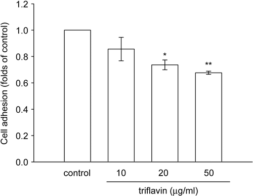
Figure 3.  Effects of triflavin on vascular smooth muscle cell (VSMC) adhesion to immobilized fibronectin. Cells (105 cell/well) were pretreated with triflavin (10, 20, and 50 μg/mL) and incubated for 1 h to allow adhesion to fibronectin-precoated wells. The number of adhering cells was evaluated as described in “Materials and methods”. Data are presented as the mean ± SEM (n = 3). *p < 0.01 and **p < 0.001, compared to the control group (PBS treatment).

Figure 4. Flow cytometric analysis of FITC–triflavin binding to vascular smooth muscle cells (VSMCs). Cells were treated with (A) FITC only or (B) FITC–triflavin (5 μg/mL); or cells were pretreated with (C) EDTA (10 mM) or (D) cold triflavin (50 μg/mL), followed by the addition of FITC–triflavin (5 μg/mL) as described in “Materials and methods”. Profiles are representative examples of three similar experiments. Data (E) are presented as folds of the control group (FITC only) and expressed as the mean ± SEM (n = 3). **p < 0.001, compared to the control group (FITC only); #p < 0.01 and ##p < 0.001, compared to the FITC–triflavin group.
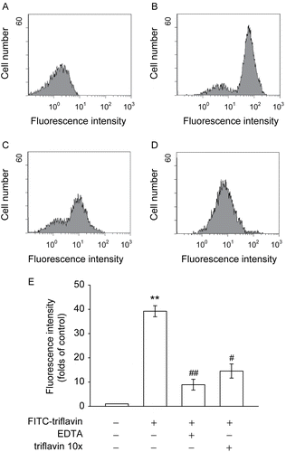
Figure 4.  Flow cytometric analysis of FITC–triflavin binding to vascular smooth muscle cells (VSMCs). Cells were treated with (A) FITC only or (B) FITC–triflavin (5 μg/mL); or cells were pretreated with (C) EDTA (10 mM) or (D) cold triflavin (50 μg/mL), followed by the addition of FITC–triflavin (5 μg/mL) as described in “Materials and methods”. Profiles are representative examples of three similar experiments. Data (E) are presented as folds of the control group (FITC only) and expressed as the mean ± SEM (n = 3). **p < 0.001, compared to the control group (FITC only); #p < 0.01 and ##p < 0.001, compared to the FITC–triflavin group.

Figure 5. Effects of triflavin on protein kinase C (PKC)-α translocation stimulated by fibronectin on vascular smooth muscle cells (VSMCs). Cells adhered to immobilized (A) poly-l-lysine (0.01%), (B) fibronectin (50 μg/mL), or (C) fibronectin (50 μg/mL) in the presence of triflavin (10 μg/mL). Confocal micrographs of the basolateral surface of cells are shown. Images are typical of those obtained in three separate experiments demonstrating the distribution of PKC-α (arrows) surrounding the cell membrane. Original magnifications ×100 (left) and ×630 (right); the bar indicates 10 μm.
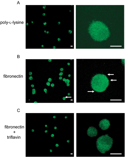
Figure 5.  Effects of triflavin on protein kinase C (PKC)-α translocation stimulated by fibronectin on vascular smooth muscle cells (VSMCs). Cells adhered to immobilized (A) poly-l-lysine (0.01%), (B) fibronectin (50 μg/mL), or (C) fibronectin (50 μg/mL) in the presence of triflavin (10 μg/mL). Confocal micrographs of the basolateral surface of cells are shown. Images are typical of those obtained in three separate experiments demonstrating the distribution of PKC-α (arrows) surrounding the cell membrane. Original magnifications ×100 (left) and ×630 (right); the bar indicates 10 μm.

Reprints and Corporate Permissions

Please note: Selecting permissions does not provide access to the full text of the article, please see our help page How do I view content?

To request a reprint or corporate permissions for this article, please click on the relevant link below:

Academic Permissions

Please note: Selecting permissions does not provide access to the full text of the article, please see our help page How do I view content?

Obtain permissions instantly via Rightslink by clicking on the button below:

If you are unable to obtain permissions via Rightslink, please complete and submit this Permissions form. For more information, please visit our Permissions help page.