Figures & data
Figure 1. (A, B) Electrocardiogram - Atrial flutter and left bundle branch block.
(C, D) Transthoracic echocardiography - Left ventricular hypertrophy (lVSd 12mm, PWd 10mm), along with speckled pattern in the myocardium. (E) Strain echocardiography - Speckled tracking revealed marked diminution of global longitudinal strain (-12) depicted in bulls-eye plot with apical sparing. (F, G) Tissue Doppler study revealed grade 2 left ventricular diastolic dysfunction (E/A mitral ratio 1,3, DT 210ms, E/è13,7). (H) Multiplanar single photon emission computed tomography (SPECT)- Showing myocardial tracer uptake, mostly in the left ventricle and septum with some heterogeneity in uptake intensity. (I) Pretreatment whole body 99mTc-3,3-diphosphono-1,2-propanodicarboxylic acid (DPD) scintigraphy - Showing very high 99mTc -DPD-uptake in the heart, Perugini grade 3.

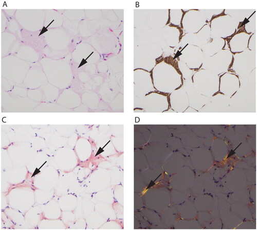
Figure 2. Fat tissue with amyloid deposition (arrows in all panels). (A) Hematoxylin eosin stain showing eosinophilic amorphous material. (B) Immunohistochemistry shows a positive reaction for transthyretin in the depositions. (C) Congo red stain is positive with orange color in the depositions and shows (D) green and orange birefringence under polarized light.