
Figures & data

Figure 1. Our study consecutively enrolled 254 patients with bifurcation lesions at the Second Hospital of Tianjin Medical University, Tianjin, China (between March 2019 and December 2022). Exclusion criteria included left main stem lesion, chronic total occlusion and the bifurcation lesions were considered unsuitable for quantitative coronary angiographic analysis according to CAG. Finally, 216 cases with non-left main bifurcation lesions were defined as the study subjects.
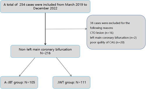
Figure 2. The procedure of the modified active jailed balloon technique. (a) Two guidewires were introduced into both the MV and the SB. (b) The MV was pre-dilated with a balloon first. The MV stent was placed to the distal of the bifurcation lesion before the jailed balloon was placed in SB. (c) The MV stent was withdrawn to an appropriate position that could cover the MV lesion, and the mid-segment of the jailed balloon was adjusted to be located at the ridge of the lesion. (d) The SB balloon was inflated, and the position of the stent was verified by angiography. (e) The MV stent was then inflated. (f) The MV stent balloon was deflated. (g) The jailed balloon was deflated. (h) The MV stent balloon was re-inflated to make the stent appose the vessel wall.
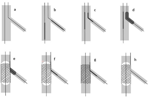
Table 1. Demographic and clinical characteristics of the study patients.
Table 2. QCA analysis of targeted lesions in the initial and final angiography.
Table 3. The characteristics of stent and balloon.
Table 4. Medications and laboratory test results during hospitalization.
Table 5. Procedural outcomes of A-JBT and JWT.
Table 6. Univariable and multivariable analyses for SB occlusion.
Figure 3. A PCI procedure was performed on a 67-year-old male patient with a bifurcation lesion in the mid-segment of the left circumflex artery. (a) The CAG displayed that was a true bifurcation of the LCX/OM. (b) The stent (3.5 mm × 20 mm) was withdrawn to an appropriate position that cover the MV lesion, and the mid-segment of the jailed balloon (1.5 mm × 16 mm) was adjusted to be located at the ridge of the lesion. (c) The MV stent was inflated at 8 atm and the jailed balloon was inflated at 11 atm. (d) The MV stent balloon was deflated. (e) The MV stent balloon was re-inflated to make the stent appose the vessel wall. (f) The diameter stenosis of the side branch had a significant improvement.
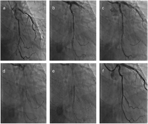
Figure 4. Diagram of bifurcation lesion.
