Figures & data
Figure 1. Effect of KN-93 on the apoptosis induced during neural differentiation with RA treatment. (A), Time course of apoptosis in the presence or absence of KN-93. Tau/P19 or wt/P19 cells were differentiated with treatment with 0.3 μM RA in the absence (left panel) or presence (right panel) of 5 μM KN-93. RA and KN-93 were added to the culture medium at 0 time, and cell viability was determined at the indicated times. (B), Effect of the concentration of differentiated cells with treatment with 0.3 μM RA in the presence or absence of the indicated amounts of KN-93 at 24 h (left panel) and 48 h (right panel) after induction of differentiation. Cell viability was determined at the indicated concentrations of KN-93.
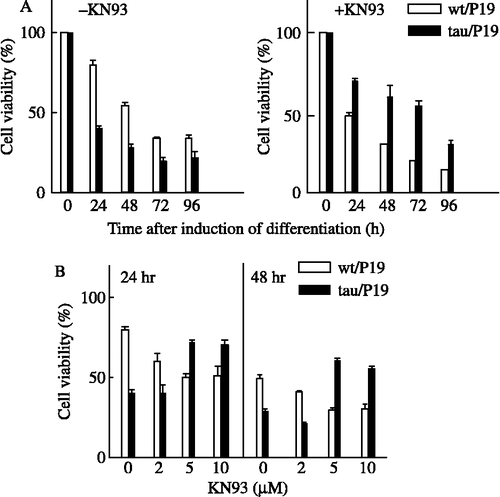
Figure 2. Effect of calmodulin antagonists or tau kinase inhibitors on apoptosis, and phosphorylation of nuclear tau at CaM kinase II site(s). (A), Effect of calmodulin antagonists or tau kinase inhibitors on the apoptosis during neural differentiation induced by RA treatment. Tau/P19 or wt/P19 cells were differentiated with treatment with 0.3 μM RA in the presence or absence of 10 μM W-7, 10 μM calmidazolium, 5 mM LiCl, and 50 nM indirubin-3′-monoxime. Cell viability was measured at 24 h after induction of differentiation. left panel, tau/P19 cells; right panel, wt/P19 cells. Abbreviations: C, control in the absence of inhibitors; Calmi, calmidazolium; Indi, Indirubin-3′-monoxime. (B), Immunoblot analysis of phosphorylation of nuclear tau at CaM kinase II site(s). Cytosolic and nuclear fractions of tau/P19 cells without RA treatment were subjected to immunoblot analysis. Each lane was applied with 50 μg of protein. Lane Tau441 was applied with purified hTau441 (100 ng). Tau was detected with H-150 antibody recognizing total tau and with 12E8 antibody recognizing phosphorylated Ser262 and Ser365. Upper panel, tau/P19 cells; lower panel, wt/P19 cells; left panel, H-150 antibody (H-150 Ab); right panel, 12E8 antibody (12E8 Ab). Abbreviations: Cyt, cytosolic fraction; Nuc, nuclear fraction.
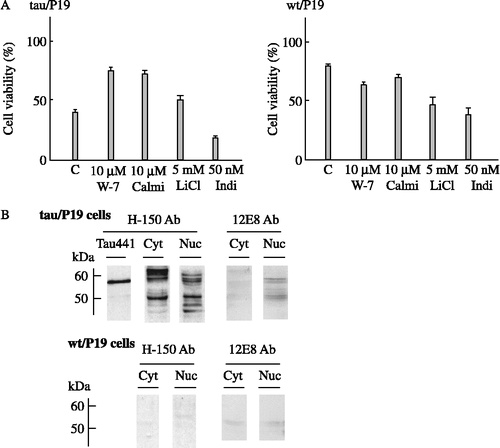
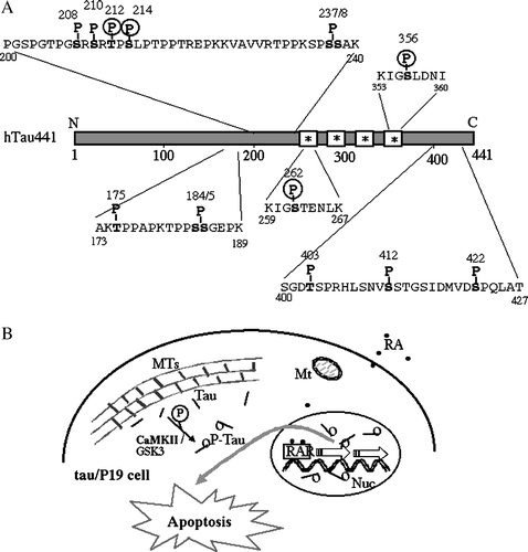
Figure 3. Schematic representation of specific phosphorylation sites of htau441 found in PHF-tau, and involvement of CaM kinase II in apoptosis of tau/P19 cells. (A), Specific phosphorylation sites of htau441 found in only PHF-tau of the AD brain. Non-fetal-type phosphorylation sites found in PHF-tau, namely PHF-tau specific phosphorylation sites, are shown, although PHF-tau is also phosphorylated at the same sites of fetal-type phosphorylation. *, tubulin binding site; P, PHF-tau-specific phosphorylation site; P in circle, phosphorylation site with CaM kinase II. (B), Involvement of CaM kinase II in apoptosis of tau/P19 cells. In tau/P19 cells, tau was distributed throughout the cytoplasm and co-localized with microtubules. Some tau dissociated from MTs was phosphorylated by CaM kinase II and/or GSK3, and then translocated into the nucleus. Nuclear tau may disrupt RA signaling during neural differentiation resulting in apoptosis. Abbreviations; MTs, microtubules; Mt, mitochondria, Nuc, nuclei; RAR, retinoic acid receptor.