
Figures & data
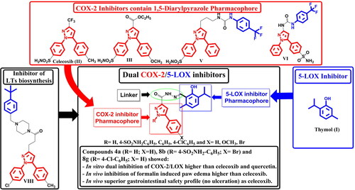
Figure 1. Structures of compounds having COX-2 or 5-LOX inhibitory activities.

Figure 2. Design of dual COX-2/5-LOX inhibitors.

Scheme 1. Synthesis of the target thymol–1,5-disubstitutedpyrazole hybrids.

Table 1. In vitro COX-1, COX-2, and 5-LOX enzyme inhibitory activities, aIC50 values, and bselectivity indices (SI) of the tested compounds.
Figure 3. Comparison between different groups according to % inhibition of paw oedema volume.

Figure 4. Comparison between the different groups according to % relative potency (4h) celecoxib.

Figure 5. Comparison between the different groups according to %relative potency (4h) diclofenac.

Table 2. In vivo anti-inflammatory activities of selected compounds in formalin-induced rat paw oedema bioassay (acute inflammation model).
Figure 6. Gross appearance of gastric mucosa.

Figure 7. Mode of binding (2D) of celecoxib (A), 4a (B), 8b (C), and 8g (D) inside the active site of COX-2.

Figure 8. Overlay of celecoxib of crystallised (yellow) and docked (cyan) with RMSD = 0.422.

Figure 9. Overlay of compounds 4a (green), 8b (yellow), 8g (pink), and celecoxib (cyan) inside the active site of COX-2.

Figure 10. Mode of binding (3D) of celecoxib (A), 4a (B), 8b (C), and 8g (D) into COX-2 active site.

Figure 11. Mode of binding (2D) of Arachidonic acid (A), 4a (B), 8b (C), and 8g (D) inside the active site of 5-LOX.

Figure 12. Overlay of compounds 4a (green), 8b (yellow), 8g (pink), and arachidonic acid (cyan) inside the active site of 5-LOX.

Figure 13. Mode of binding (3D) of arachidonic acid (A), 4a (B), 8b (C), and 8g (D) inside the active site of 5-LOX.

Table 3. Docking results of the active compounds in COX-2 active site.
Table 4. Docking results of the active compounds in 5-LOX active site.