Figures & data
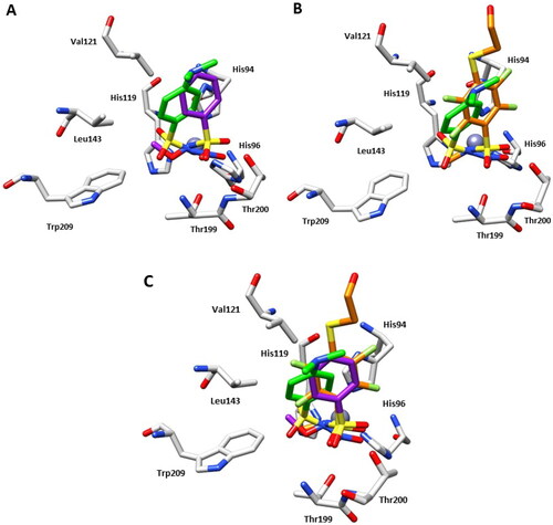
Figure 1. Structures of benzenesulfonamide 1 and N-substituted sulphonamides 2–5.
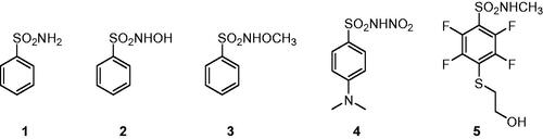
Scheme 1. Synthetic route to obtain the N-nitrosulfonamide derivative 4.
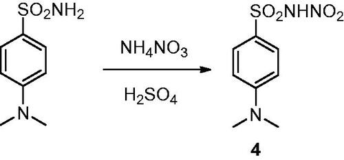
Table 1. Inhibition data of human CA isoforms I, II, IV, IX, and XII with compounds 1–4 and AAZCitation17,Citation19,Citation20.
Figure 2. (A) Active site region of the 4-hCA II complex (PDB: 8BZZ). Hydrogen bonds (red), van der Waals interactions (blue), and zinc interactions (green) are also shown. (B) Structural superposition between 4-hCA II (green) and 2-hCA II (magenta, PDB: 3T5U)Citation17 bound to the active site of protein. (C) Structural superposition between 4-hCA II (green) and 1-hCA II (cyan, PDB: 2WEJ)Citation21 bound to the active site of protein.
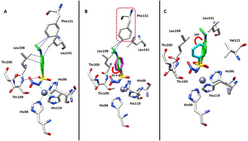
Figure 3. (A) Structural superposition between 4-hCA II (green) and 3-hCA II (purple, PDB: 3T5Z)Citation17 bound to the active site of protein. (B) Structural superposition between 4-hCA II (green) and 5-hCA II (orange, PDB: 7AEQ)Citation23 bound to the active site of protein. (C) Structural superposition between 4-hCA II (green), 3-hCA II (purple), and 5-hCA II (orange) bound to the active site of protein.