Figures & data
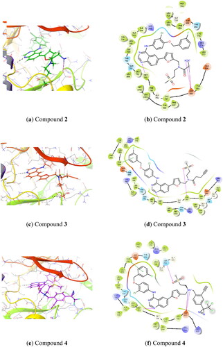
Figure 1. Predicted binding of lapatinib analogues to the EGFR receptor site (docked to PDB: 1xkk) (1a, 1c, 1e) and corresponding schematic diagrams (1b, 1d, 1f).
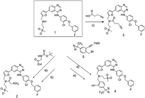
Scheme 1. Synthetic approach to synthesised analogues of lapatinib. Reagents and conditions: (a) PS-CDI, HOBT, DIPEA, DCM, DMF, rt; (b) TFA, DCM, rt; (c) PS-CDI, HOBT, DIPEA, DCM, DMF, rt; (d) NaH, DMF, rt; (e) K2CO3, MeOH, rt.
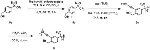
Scheme 2. Synthesis of intermediate 5. Reagents and conditions: (a) thallium(III) trifluoroacetate, TFA, NaI, CF3SO3H, H2O, 80 °C, 2h; (b) (trimethylsilyl)acetylene, CuI, TEA, PdCl2(PPh3)2, THF, rt, on; (c) Ph3P, CBr4, DCM, rt, on.
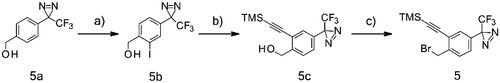
Table 1. In vitro activity of lapatinib and its analogues as EGFR inhibitors.
Figure 2. Silver-stained gels after interaction of purified recombinant EGFR with immobilised lapatinib derivatives 3 (a) and 2 (b). Matrices alone were used as negative controls (/). Purified recombinant EGFR was used as additional control.
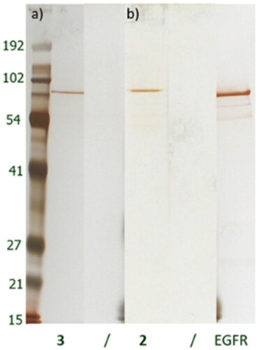
Figure 3. Silver stained gels of bound proteins after incubation of 2 (a) and 3 (b) derivatives with A431 cell lysate.
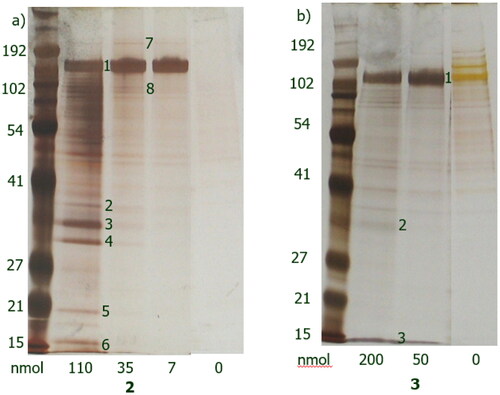
Table 2. Identified proteins captured by 2, as identified using ESI-MS.
Table 3. Identified proteins captured by 3, as identified using ESI-MS.
Table 4. Identified proteins captured by 4, as identified using ESI-MS.
Table 5. Identified lapatinib bound proteins across different proteomics technologies.
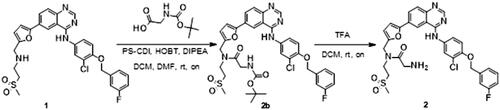
Scheme 3. Synthesis of analogue 2. Reagents and conditions: (a) PS-CDI, HOBT, DIPEA, DCM, DMF, rt; (b) TFA, DCM, rt.
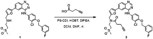
Scheme 4. Synthesis of analogue 3. Reagents and conditions: (a) PS-CDI, HOBT, DIPEA, DCM, DMF, rt.
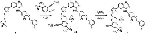
Scheme 5. Synthesis of analogue 4. Reagents and conditions: (a) NaH, DMF, rt; (b) K2CO3, MeOH, rt.
Scheme 6. Synthesis of intermediate 5. Reagents and conditions: (a) thallium(III) trifluoroacetate, TFA, NaI, CF3SO3H, H2O, 80 °C, 2h; (b) (trimethylsilyl)acetylene, CuI, TEA, PdCl2(PPh3)2, THF, rt, on; (c) Ph3P, CBr4, DCM, rt, on.