1,088
Views
0
CrossRef citations to date
0
Altmetric
Research Article

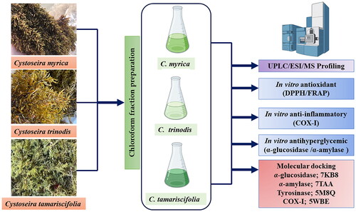
Comparative metabolic study of the chloroform fraction of three Cystoseira species based on UPLC/ESI/MS analysis and biological activities

ORCID Icon, , , , , , ORCID Icon & ORCID Icon show all
Article: 2292482 | Received 07 Oct 2023, Accepted 04 Dec 2023, Published online: 12 Dec 2023

Figures & data

Table 1. Metabolite profiling of C. myrica, C. trinodis and C. tamariscifolia Chloroform fraction using UPLC-ESI-MS in the negative ion mode.

Figure 1. Chemical structures of the major characterised metabolites in three Cystoseira species using UPLC-ESI-MS in the negative ion mode.

Figure 1. Chemical structures of the major characterised metabolites in three Cystoseira species using UPLC-ESI-MS in the negative ion mode.

Table 2. Total phenolic and flavonoid content of C. myrica, C. trinodis and C. tamariscifolia chloroform fraction.

Table 3. Antioxidant activity (IC50) of C. myrica, C. trinodis and C. tamariscifolia chloroform fraction using DPPH and FRAP assays.

Table 4. Anti-hyperglycaemic activity and Anti-inflammatory activity % (IC50) of C. myrica, C. trinodis and C. tamariscifolia chloroform fraction.

Table 5. The docking scores achieved by the major identified compounds towards various enzymes.

Figure 2. (I) 2D binding modes of (A) 4′,7-Dimethoxyluteolin, (B) 2″,3″-Dihydro-3',3'″-biapigenin methyl ether, (C) Naringin to the active binding sites of α-amylase enzyme. (II) 2D binding modes of (A) 4′,7-dimethoxyluteolin, (B) 2″,3″-dihydro-3',3'″-biapigenin methyl ether, (C) Naringin to the active binding sites of α-glucosidase enzyme.

Figure 2. (I) 2D binding modes of (A) 4′,7-Dimethoxyluteolin, (B) 2″,3″-Dihydro-3',3'″-biapigenin methyl ether, (C) Naringin to the active binding sites of α-amylase enzyme. (II) 2D binding modes of (A) 4′,7-dimethoxyluteolin, (B) 2″,3″-dihydro-3',3'″-biapigenin methyl ether, (C) Naringin to the active binding sites of α-glucosidase enzyme.

Figure 3. (I) 2D binding modes of (A) 4',7-Dimethoxyluteolin, (B) 2'',3''-Dihydro-3',3'''-biapigenin methyl ether, (C) Naringin to the active binding sites of COX-1 enzyme. (II) 2D binding modes of (A) Cystophloroketal B, (B) 2'',3''-Dihydro-3',3'''-biapigenin methyl ether, (C) Quercetin to the active binding sites of Tyrosinase enzyme.

Figure 3. (I) 2D binding modes of (A) 4',7-Dimethoxyluteolin, (B) 2'',3''-Dihydro-3',3'''-biapigenin methyl ether, (C) Naringin to the active binding sites of COX-1 enzyme. (II) 2D binding modes of (A) Cystophloroketal B, (B) 2'',3''-Dihydro-3',3'''-biapigenin methyl ether, (C) Quercetin to the active binding sites of Tyrosinase enzyme.
Supplemental material

Supplemental Material

Download PDF (584.6 KB)