
Figures & data
Figure 1. Consecutive enzymatic processing steps of MSP1 protein. MSP1 is produced at the end of schizogony as a precursor protein of around 196 kDa. Once the merozoite reaches maturity, PfSUB1 cleaves MSP1 at specific cleavage sites (shown here in gray squares), leading to formation of four fragments named p83 (MSP1-83), p30 (MSP1-30), p38 (MSP1-38), and p42 (MSP1-42). A second cleavage event of the p42 fragment by PfSUB2 leads to the formation of fragments p33 (MSP1-33) and p19 (MSP1-19). MSP1 is divided in 17 domains [Citation37] based on sequence polymorphisms, whereby 7 domains are highly polymorphic, 5 are semi-conserved, and 5 are highly conserved which are color-marked in red, orange, and pink, respectively. MSP1 has been historically characterized by two prototypic sequences: the MSP1-D from the P. falciparum MAD20 strain and the MSP1-F from the WELLCOME strain. These two forms are found in African parasite populations, with distinct distributions observed between East and West Africa [Citation36,Citation148].
![Figure 1. Consecutive enzymatic processing steps of MSP1 protein. MSP1 is produced at the end of schizogony as a precursor protein of around 196 kDa. Once the merozoite reaches maturity, PfSUB1 cleaves MSP1 at specific cleavage sites (shown here in gray squares), leading to formation of four fragments named p83 (MSP1-83), p30 (MSP1-30), p38 (MSP1-38), and p42 (MSP1-42). A second cleavage event of the p42 fragment by PfSUB2 leads to the formation of fragments p33 (MSP1-33) and p19 (MSP1-19). MSP1 is divided in 17 domains [Citation37] based on sequence polymorphisms, whereby 7 domains are highly polymorphic, 5 are semi-conserved, and 5 are highly conserved which are color-marked in red, orange, and pink, respectively. MSP1 has been historically characterized by two prototypic sequences: the MSP1-D from the P. falciparum MAD20 strain and the MSP1-F from the WELLCOME strain. These two forms are found in African parasite populations, with distinct distributions observed between East and West Africa [Citation36,Citation148].](/cms/asset/b7f68b54-b46f-4473-b5bd-0a63e8b650a4/ierv_a_2295430_f0001_oc.jpg)
Figure 2. Genetic diversity of MSP1 field isolates. (A) UMAP single cell expression profile of the merozoite surface protein gene PF3D7_0930300; https://www.sanger.ac.uk/tool/mca/mca/. Each dot represents a cell. The four quadrants represent the transcriptomes of sporozoites (salivary glands, top left quadrant), sporozoites (hemolymph, lower left quadrant), and ookinetes (lower right quadrant). The top right quadrant, containing the cells in which MSP1 is most highly expressed, is asexual blood-stage parasites. (B) Geographic origin of P. falciparum strains from which MSP1 sequences were obtained and frequency of representative alleles based on block 2 classification. Lower rows (in gray) are stratified African samples according to WHO regional delineations. (C) Principal component analysis (PCA) of msp1 sequences aligned with MUSCLE in MegaX v10.0.5. PCA values were calculated using multi-sequence alignment with Rv.4.1.3 library adegenet.
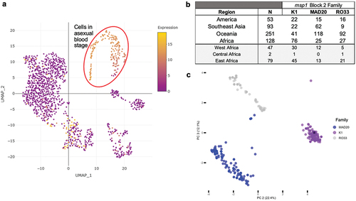
Figure 3. Overview of MSP1-based vaccine constructs evaluated in preclinical and early phase clinical development studies. Arrows point at the MSP1 subunit or epitopes within the subunit contained in the vaccine candidate. Highlighted: block 2, dimorphic fragments M6 (amino acids 671–833 from the K1 strain in yellow) and F5 (amino acids 384–595 from the MAD20 strain in orange).
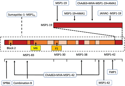
Table 1. Subunit and viral vectored MSP1 vaccine constructs evaluated in clinical trials.
Supplemental Material
Download Zip (605.3 KB)Data availability statement
The Plasmodium falciparum whole-genome assemblies used for this work are available from the NCBI (https://www.ncbi.nlm.nih.gov/gene/).