1,816
Views
3
CrossRef citations to date
0
Altmetric
Research Article

Prenatal ultrasound evaluation of fetal cutaneous hemangioma and related complications

, , , , , , & show all
Article: 2157257 | Received 16 Sep 2022, Accepted 02 Dec 2022, Published online: 15 Dec 2022

Figures & data

Figure 1. A: (case 17) cervical cutaneous hemangioma at 27 weeks gestation. A1, a mixed mass mainly in the front neck, with a size of about 56 × 33 × 56 mm and internal tubular anechoic signals (arrow). A2 shows abundant blood flow signals within the mass by the color Doppler (arrow). B: (case 5) B1, a mixed echogenic mass was seen in the left shoulder, left axillary area, and left chest wall, with a size of about 58 × 41 × 40 mm, with internal honeycomb echoes (arrow). B2, the rich blood flow signal can be detected internally, with burr-like blood flow spectrum. B3, unenhanced MRI SSFSE sequence shows multiple tortuous vessels with low signal intensities (arrow). C: (case 1) C1, at 23 weeks of gestation, a solid uniform echogenic mass, with a size of 22 × 17 × 19 mm and clear borders, was detected in the dorsal side of right lower leg (arrow). C2, three-dimensional reconstruction (arrow). C3, after birth. C4, surgical resected specimens (arrow). D: (case 12) D1, a hyperechoic well-circumscribed mass in the precordial area of the left chest wall, about 27 × 18 × 33 mm and regular shape (arrow). D2, three-dimensional reconstruction (arrow). E: (case 14) at 35 weeks of gestation, hemangioma from the right buccal extending to the frontal chest wall. E1, internal blood supply is shown in Doppler images (arrow). E2, three-dimensional reconstruction (arrow). E3, unenhanced MRI FIESTA sequence shows a mass (triangle arrow) compressing the esophagus (large arrow) and traches (small arrow). F: (case 2) At 30 weeks of gestation. F1, a cutaneous hemangioma was observed on the top of the head (arrow). F2, the internal structure of the mass was clearly shown by a high frequency line array probe (arrow).

Figure 1. A: (case 17) cervical cutaneous hemangioma at 27 weeks gestation. A1, a mixed mass mainly in the front neck, with a size of about 56 × 33 × 56 mm and internal tubular anechoic signals (arrow). A2 shows abundant blood flow signals within the mass by the color Doppler (arrow). B: (case 5) B1, a mixed echogenic mass was seen in the left shoulder, left axillary area, and left chest wall, with a size of about 58 × 41 × 40 mm, with internal honeycomb echoes (arrow). B2, the rich blood flow signal can be detected internally, with burr-like blood flow spectrum. B3, unenhanced MRI SSFSE sequence shows multiple tortuous vessels with low signal intensities (arrow). C: (case 1) C1, at 23 weeks of gestation, a solid uniform echogenic mass, with a size of 22 × 17 × 19 mm and clear borders, was detected in the dorsal side of right lower leg (arrow). C2, three-dimensional reconstruction (arrow). C3, after birth. C4, surgical resected specimens (arrow). D: (case 12) D1, a hyperechoic well-circumscribed mass in the precordial area of the left chest wall, about 27 × 18 × 33 mm and regular shape (arrow). D2, three-dimensional reconstruction (arrow). E: (case 14) at 35 weeks of gestation, hemangioma from the right buccal extending to the frontal chest wall. E1, internal blood supply is shown in Doppler images (arrow). E2, three-dimensional reconstruction (arrow). E3, unenhanced MRI FIESTA sequence shows a mass (triangle arrow) compressing the esophagus (large arrow) and traches (small arrow). F: (case 2) At 30 weeks of gestation. F1, a cutaneous hemangioma was observed on the top of the head (arrow). F2, the internal structure of the mass was clearly shown by a high frequency line array probe (arrow).

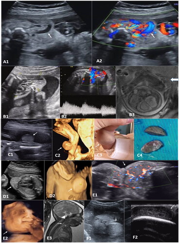
Table 1. Size and growth rate of fetal cutaneous hemangioma diagnosed at different gestational weeks in 15 cases who chose to continue pregnancy.

Table 2. Prenatal ultrasound findings and pregnancy outcomes in 8 fetuses with complications.

Supplemental material

Supplemental Material

Download MS Word (16.6 KB)