
Figures & data
Figure 1. A scheme showing the basic steps for intrasurgical PAS staging. Left: After cutting the peritoneum medially to the round ligament (RL), fingers open the parametrial space between avascular fascia sheets. Right: From the upper position (A), the index goes down (B) by the subperitoneal tissue until crossing the fingers behind the bladder (BL) to perform a Pelosi maneuver. IA: invaded area.
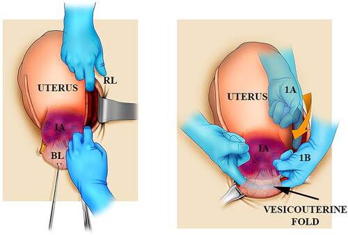
Figure 2. Parasagittal cut on the right female pelvis. An embalmed corpse, the peritoneal sheet of the parametrium was transilluminated. Notice that the parametrium in a straight space has plenty of arteries, veins, and the ureter.
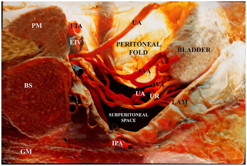
Table 1. Clinical results of patients with upper and lower type 2 PAS placenta invasion.
Supplemental material