
Figures & data
Figure 1. Flowchart included and excluded cases. TOP: termination of pregnancy; CNS: central nervous system; PMMR: postmortem MR; PI: Pathological investigations. Genetic anomaly: chromosomal anomaly + anomaly detectable by array comparative genomic hybridization (CGH).
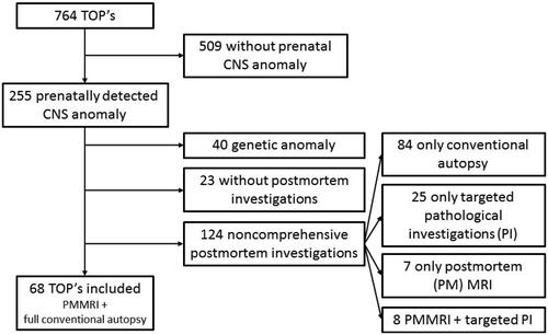
Table 1. Conventional autopsy provides more information than postmortem.
Table 2. Cases in which PM MR detects major additional findings, undetected prenatally and undetected by CA, influencing final diagnosis and parental counseling.
Figure 2. Distribution of detected CNS anomalies. X-axis = number of cases. ISV = isolated severe ventriculomegaly; NTD = neural tube defect.
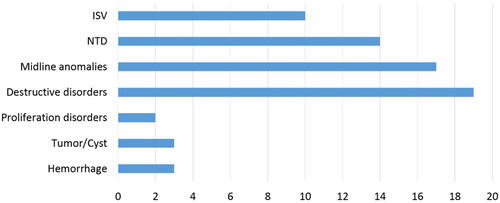
Figure 3. Case: Prenatal diagnosis of hydrocephalus, cerebellar hypoplasia and aqueductal stenosis. TOP at GA 27 weeks. PM MR shows hydrocephalus, rhombencephalosynapsis, CC dysgenesis, vertebral anomalies. CA shows hydrocephalus, rhombencephalosynapsis, CC dysgenesis. Vertebral anomalies are only depicted on PM MR, demonstrating superiority of PM MR over CA in this case. (a) T2-weighted image of the brain in the axial plane. Dilated right temporal horn of the lateral ventricle (black star). Black fluid surrounding the cerebellum (white arrows). The dentate nuclei appear fused (White circle) and there is no vermian structure visible (curved white line). (b) T2-weighted image of the brain in the axial plane. Severe ventriculomegaly with blood layering as a normal post mortem finding and destruction of the leaflets of the cavum septi pellucidi (thin black arrow). Furthermore, the normal layering of the cerebral mantle is seen with the germinative matrix, subventricular zone, subplate and cortical plate. (c) T1-weighted image of the body in the coronal plane. Abnormal ossification of a thoracic vertebral body (T11) in the lower half of the thoracic spine. (d) T1-weighted image of the body in the coronal plane. Abnormal ossification of two thoracic vertebral bodies (T1 and T4) in the upper half of the thoracic spine.
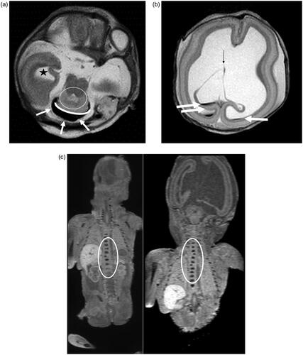
Table 3. The time interval between feticide, delivery and PM MR in different groups with and without feticide or autolysis.