
Figures & data
Figure 1. Echocardiographic appearance of the mass at 23 weeks of gestational age.
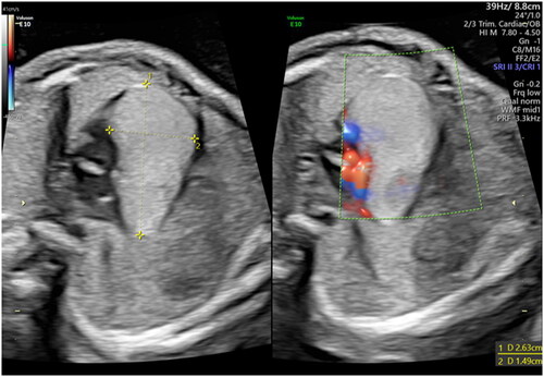
Figure 2. Echocardiographic appearance of the rhabdomyoma at 25 (a) and 37 (b) weeks of gestational age respectively, demonstrating its progressive enlargement.
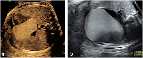
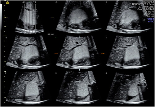
Figure 3. Three-dimensional reconstruction of the rhabdomyoma at 37 weeks by Tomographic Ultrasound Imaging (TUI).
Supplemental material