
Figures & data
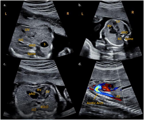
Figure 1. Fetal echocardiography performed at 22w1d. (a) Abdominal situs view. The stomach (S) is localized on the right side of the fetus. In front of and slightly to the left of the spine is the descending aorta (dAO). The umbilical vein (UV) and the inferior vena cava (IVC) are present anteriorly and to the left. (b) Four chamber view. The dAO and the azygos vein (AZ, further to the left) are anterior to the spine. RV: right ventricle; LV: left ventricle. (c) Transverse view. RV: right ventricle; LV: left ventricle; aAO: ascending aorta. The dAO and the AZ (to the left) are anterior to the spine. (d) Sagittal aortic arch and azygos vein view. Inferior to the flow of the aortic arch a red flow is visible, attributable to the azygos continuation of the inferior vena cava.
Data availability statement
The data that support the findings of this study are available from the corresponding author upon reasonable request.