Figures & data
Table 1 Patient demographics of the population studied (data expressed as median ± data range), * p < 0.05 versus never smoker controls
Table 2 Mabs used for investigation of chemokine receptor expression
Figure 1 Representative flow cytometry dot plots of chemokine receptor expression on T-cells from blood (A-E) and BAL (F-G) from a never smoker control and a COPD subject. A. and F. CCR5; B and G. CXCR3; C. CCR4; D. CXCR4 and E. intracellular CCR3. T cells were selected using SSC vs. CD3 (data not shown). Quadrant markers were set using a cell population known to lack expression of the marker of interest. The chemokine [PE stained] of interest is on the y-axis and CD8 [FITC] on the x-axis (with the exception of CCR5 [FITC] on the x-axis with CD8 [PE] on the y-axis). The percentage of cells expressing chemokine receptors are shown in the relevant quadrants.
![Figure 1 Representative flow cytometry dot plots of chemokine receptor expression on T-cells from blood (A-E) and BAL (F-G) from a never smoker control and a COPD subject. A. and F. CCR5; B and G. CXCR3; C. CCR4; D. CXCR4 and E. intracellular CCR3. T cells were selected using SSC vs. CD3 (data not shown). Quadrant markers were set using a cell population known to lack expression of the marker of interest. The chemokine [PE stained] of interest is on the y-axis and CD8 [FITC] on the x-axis (with the exception of CCR5 [FITC] on the x-axis with CD8 [PE] on the y-axis). The percentage of cells expressing chemokine receptors are shown in the relevant quadrants.](/cms/asset/4f40f4c0-bf4d-43b3-b38a-0f4ce8ef85ff/icop_a_372586_uf0001_b.gif)
Table 3 Differential cell counts
Table 4 Summary of results
Figure 2 A. Cell membrane expression of CCR5 and CXCR3 (as a percentage of CD3+ T-cells) in peripheral blood (mean ± SEM). Similar results were noted for CD8+ T-cells (not presented). B. BAL. Cell membrane expression of CCR5 and CXCR3 (as a percentage of CD3+ T-cells). * p < 0.05 versus never-smoker controls.
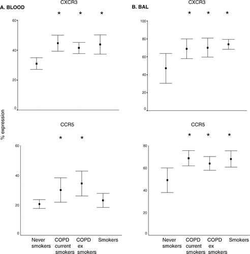
Figure 3 Intracellular expression of CCR3 and cell membrane expression of CCR4 (as a percentage of CD3+ T-cells) in peripheral blood (mean ± SEM). * p < 0.05 versus never smoker controls. Note increased expression of CCR4 and intracellular CCR3 in all groups compared to never smoker controls. Similar results were noted for CD3+ T-cells (not presented). Data is presented as box plots showing median ± 25th and 75th percentiles (solid box) and the 10th and 90th percentiles shown by whiskers outside the box.
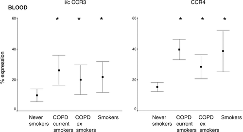
Figure 4 Cytotoxic and Th1 markers in A. peripheral blood and B. BAL (mean ± SEM). Th1 type T-cells were considered to be CD3+CD45RO+CD62L− T-cells. Cytotoxic T-cells were considered to be CD8+CD11abright or CD8+CD28bright. * p < 0.05 versus never smokers. Note the significantly increased percentage of Th1 type T-cells in both COPD groups and healthy smokers but an increase in cytotoxic T-cells only in the COPD groups.
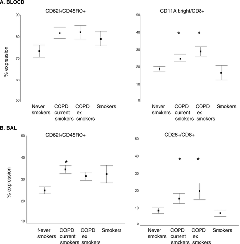
Figure 5 Intracellular expression of IFN-γ (as a percentage of CD8+ T-cells) in A. blood and B. BAL from COPD subjects, asymptomatic smokers and healthy never-smokers measured current and ex-smoker by flow cytometry (mean ± SEM). * p < 0.05 versus never-smokers. There were significantly increased levels of IFN-γ in blood CD8+ T-cells from both current and ex-smoker COPD subjects but no change in healthy smokers compared to never-smoker controls.
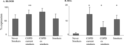
Figure 6 Soluble MCP-1, MIG and ITAC levels in plasma and BAL from current and ex-smoker COPD subjects, healthy smokers and never-smoker controls (mean ± SEM). MCP-1 and ITAC were measured by ELISA and MIG by cytometric bead array A. plasma B. BAL. * p < 0.05 versus never-smokers. All chemokines were increased in BAL from all groups compared to controls. Note increased levels of MIG and ITAC in plasma from both current and ex-smoker COPD subjects but no change in healthy smokers.
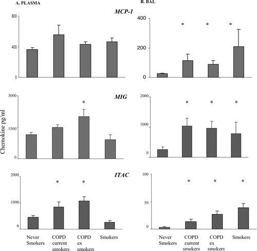
Figure 7 Soluble cytokine levels in BAL from current and ex-smoker COPD subjects, healthy smokers and never-smoker controls measured by cytometric bead array (mean ± SEM). A. RANTES B. IL-8. * p < 0.05 versus never-smokers. ** p < 0.05 versus current smokers. ns: no significant differences between groups. There were no significant differences in levels of these soluble chemokines in plasma (not shown).
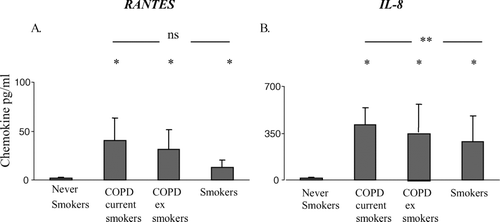
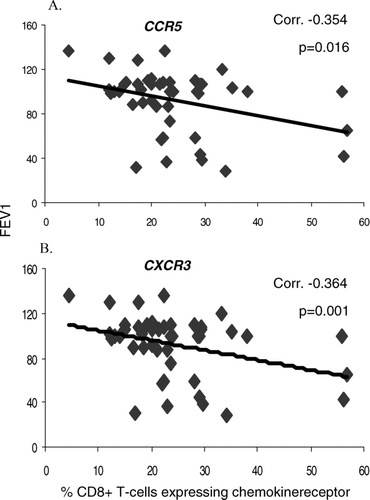
Figure 8 Correlation between A. % CCR5 expression and B. % CXCR3 expression by peripheral blood CD8+ T-cells and COPD disease severity (FEV1).