Figures & data
Figure 1 Diagram of the DNA construct/system use in stable transfection. (A) Constitutive CMV promoter constructs (native and tagged versions, three different antibiotic resistances cloned downstream from an internal ribosome entry site (IRES) are also available) were transfected into HeLa cells. Lysates from stable transfectants expressing Cx40, Cx43, Cx45, and the tagged versions of these connexins were used to calibrate the specific anti-connexin antibodies' reactivity in Western blot experiments and thereby to compare the connexins to each other. The position of the V5 epitope and the six-histidine stretch are indicated. (B) Diagram of the ecdysone system that permits inducible expression of Cx40 and Cx45. RLE cells were first transfected with pVgRXR (Invitrogen) coding for the transcription factors of the ecdysone system (ecdysone receptor, VgEcR; retinoid X receptor [RXR]) and can be selected using bleomycin. One stable clone expressing the transcription factors was retransfected with DNA constructs coding for both a connexin (CxX; Cx40 or Cx45) and the hygromycin resistant gene (Hygro) downstream from the IRES on the bicistronic construct. Upon association with ponasterone A, the VgEcR and RXR proteins form a heterodimer that activates the IND promoter and induces the transcription of an mRNA coding for Cx40 or Cx45 and hygromycin resistance. Selection of inducible RLE clones was therefore done in induction medium since the uninduced cells are not resistant to the antibiotic.
![Figure 1 Diagram of the DNA construct/system use in stable transfection. (A) Constitutive CMV promoter constructs (native and tagged versions, three different antibiotic resistances cloned downstream from an internal ribosome entry site (IRES) are also available) were transfected into HeLa cells. Lysates from stable transfectants expressing Cx40, Cx43, Cx45, and the tagged versions of these connexins were used to calibrate the specific anti-connexin antibodies' reactivity in Western blot experiments and thereby to compare the connexins to each other. The position of the V5 epitope and the six-histidine stretch are indicated. (B) Diagram of the ecdysone system that permits inducible expression of Cx40 and Cx45. RLE cells were first transfected with pVgRXR (Invitrogen) coding for the transcription factors of the ecdysone system (ecdysone receptor, VgEcR; retinoid X receptor [RXR]) and can be selected using bleomycin. One stable clone expressing the transcription factors was retransfected with DNA constructs coding for both a connexin (CxX; Cx40 or Cx45) and the hygromycin resistant gene (Hygro) downstream from the IRES on the bicistronic construct. Upon association with ponasterone A, the VgEcR and RXR proteins form a heterodimer that activates the IND promoter and induces the transcription of an mRNA coding for Cx40 or Cx45 and hygromycin resistance. Selection of inducible RLE clones was therefore done in induction medium since the uninduced cells are not resistant to the antibiotic.](/cms/asset/9254dd97-5c42-4883-ac46-da59b2531cfb/icac_a_301560_uf0001_b.gif)
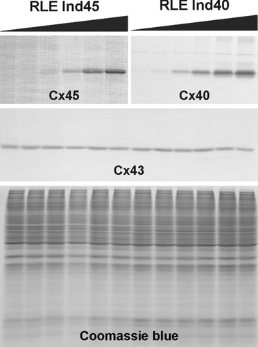
Figure 2 Dose-dependent expression of transfected connexins upon induction with ponasterone A. The upper immunoblots show the induction of Cx45 or Cx40 in the RLE Ind45 and RLE Ind40 cell lines, respectively. The black gradient slopes indicate increasing ponasterone A concentrations (0, 0.1, 0.25, 0.5, 1, and 2 μ M). Expression of both Cx45 and Cx40 is tightly regulated by the inducer. Endogenous Cx43 expression (detected with the anti-Cx43 Sigma antibody) is shown in the middle panel and equal protein loading is demonstrated by Coomassie blue staining (lower panel).

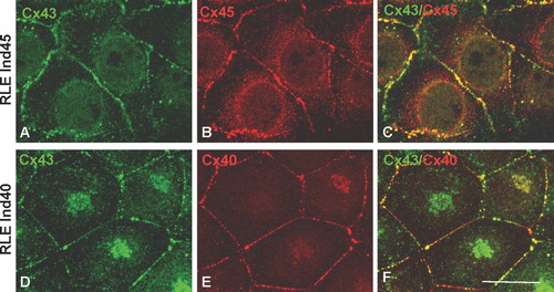
Figure 3 Double immunolabeling of Cx43 and Cx45 in RLE Ind45 cells (A, B, C) and Cx43 and Cx40 in RLE Ind40 cells (D, E, F) at maximal induction. Cx43 (detected using the Chemicon antibody) is colocalized with Cx45 or Cx40 in the same junctions. Scale bar: 25 μ m.

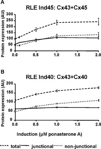
Figure 4 Quantitative analysis of immunoblots to determine total, nonjunctional and junctional connexins in transfected RLE cell lines. (A) In RLE Ind45 cells, total connexin levels are increased by a factor of 2.4 at maximum induction. However, junctional Cx43/Cx45 is increased only by a factor of 2. (B) In RLE Ind40 cells, total connexin levels are increased by a factor of 1.7 at maximum induction, but junctional Cx40/Cx43 remains constant. 100 AU is defined as the total amount of endogenous Cx43 in both cell lines in the noninduced state.

TABLE 1 Proportion of nonjunctional and junctional Cx43 (endogenous) and Cx45 with increasing levels of induction
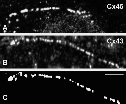
Figure 5 Representative images used to measure gap junction size. In this example, RLE Ind45 cells at maximal induction were labeled for either Cx45 (A) or Cx43 (B; Chemicon). Images were thresholded before measuring gap junction size with PC Image software (C). Scale bar: 5 μ m.

Figure 6 Gap junction sizes measured in RLE Ind45 cells at varying levels of induction. In order to compare gap junction size labeled with the anti-Cx43 and anti-Cx45 antibodies, data were normalized to the average size at maximum induction (100 arbitrary units [AU]). At both medium and high levels of induction Cx43-labeled gap junction size (measured with both the Chemicon and Sigma antibodies) was significantly reduced compared to noninduced cells. The size of Cx45-labeled gap junctions in induced cells was comparable to those measured for Cx43. The concentrations (0, 0.5, 2 μ M) of ponasterone A used are indicated. * p < 0.001.
![Figure 6 Gap junction sizes measured in RLE Ind45 cells at varying levels of induction. In order to compare gap junction size labeled with the anti-Cx43 and anti-Cx45 antibodies, data were normalized to the average size at maximum induction (100 arbitrary units [AU]). At both medium and high levels of induction Cx43-labeled gap junction size (measured with both the Chemicon and Sigma antibodies) was significantly reduced compared to noninduced cells. The size of Cx45-labeled gap junctions in induced cells was comparable to those measured for Cx43. The concentrations (0, 0.5, 2 μ M) of ponasterone A used are indicated. * p < 0.001.](/cms/asset/791a4344-583d-4b06-8af3-473930834c1a/icac_a_301560_uf0006_b.gif)
Figure 7 Anti-Cx43 antibody (Sigma) cross-reacts with Cx40. Screening of Cx40 transfected HeLa cells using (A) anti-Cx40 (S15C(R84)), (B) anti-Cx43 (Sigma), and (C) anti-Cx43 (Chemicon). Cx40 is detected at cell interfaces (A). Labeling is also seen at cell interfaces when using the anti-Cx43 antibody (Sigma) (B). However, labeling with anti-Cx43 antibody (Chemicon) indicates that Cx43 is not expressed and the apparent anti-Cx43 Sigma antibody labeling is due to cross-reactivity with Cx40 (C). Scale bar: 100 μ m.
