Figures & data
TABLE 1 Thymocyte and splenocyte subpopulation percentages and cell counts after SMD exposure
FIG. 1 Dose-response effect of SMD on thymic and splenic cellularity. Mice were administered SMD orally at 9:00 PM daily for 3 days. On day 4 thymi (a) and spleens (b) were collected and cellularity was determined. N = 5 mice per group. Asterisks (*) indicate significant difference from naïve control (p < 0.05).
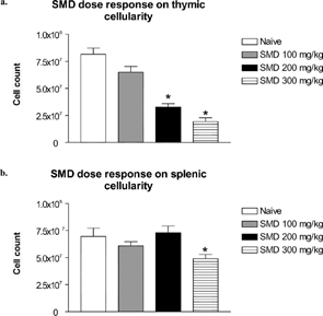
FIG. 2 SMD-increased serum corticosterone in mice. Mice were administered SMD (300 mg/kg) orally at 9:00 PM. After 1, 2, 4, 6, or 8 hours, trunk blood was collected and serum was isolated for corticosterone analysis by RIA. N = 5 mice per group. Asterisks (*) indicate significant difference from naïve control (p < 0.05).
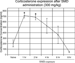
FIG. 3 SMD did not cause thymic atrophy in adrenalectomized mice. Adrenalectomized mice were administered SMD (200 mg/kg) orally at 9:00 PM on day 1 only. On day 3, thymi and spleens were removed and analyzed for alterations in cellularity. Corticosterone levels were analyzed for all samples. Two samples (each from a different group) were omitted due to the presence of serum corticosterone, indicative of adrenal regeneration. N = 5 mice per group. Asterisks (*) indicate significant difference from naïve control (p < 0.05).
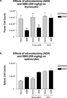
FIG. 4 Aminoglutethimide blocked serum corticosterone for up to 8 hours in vivo. Mice were administered aminoglutethimide for 8, 4, and 1 hour. One hour before collection of serum, all mice (except naïve) were restrained to induce a stress response. Trunk blood was collected at 9:00 AM and serum was isolated for analysis of corticosterone by RIA. N = 5 mice per group. Asterisks (*) indicate significant difference from naïve control (p < 0.05).
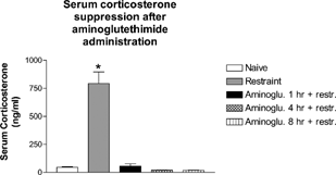
FIG. 5 SMD did not cause thymic atrophy in combination with aminoglutethimide. Mice were pretreated with aminoglutethimide 1 hour before SMD administration. SMD was administered (200 mg/kg) daily for 3 days. On day 4, thymi (a) and spleens (b) were collected and analyzed for alterations in cellularity. N= 5 mice per group. Asterisks (*) indicate significant difference from naïve control (p < 0.05).
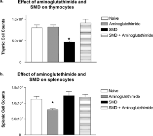
TABLE 2 Aminoglutethimide blocks alterations in thymic subpopulations caused by SMD
FIG. 6 Aminoglutethimide blocked serum corticosterone up-regulation by SMD. Mice were pretreated with aminoglutethimide for 1 hour before SMD administration. SMD was administered (200 mg/kg) by oral gavage. After 1 hour, trunk blood was collected and serum was analyzed by RIA for serum corticosterone. N = 5 per group. Asterisks (*) indicate significant difference from naïve control (p < 0.05).
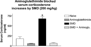
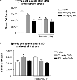
FIG. 7 SMD intoxication at a high and low dosage in combination with a psychogenic/neurogenic stressor was not synergistic. Mice were dosed with SMD (200 or 50 mg/kg) at 9:00 PM for 3 consecutive days. Immediately after SMD dosage, mice were restrained for 2 hours in their home cages. On day 4, thymi and spleens were removed and analyzed for alterations in cellularity. N = 5 per group. Asterisks (*) indicate significant difference from naïve control (p < 0.05).