523
Views
9
CrossRef citations to date
0
Altmetric
Research Article

Use of Avian Lymphocytes to Detect Toxicity: Effects of a Commonly Utilized Deltamethrin Preparation

, &
Pages 101-109 | Received 26 Dec 2005, Accepted 03 Apr 2006, Published online: 09 Oct 2008

Figures & data

TABLE 1 In vitro effect of deltamethrin on B- and T-lymphocyte proliferation in the presence LPS or Con-A mitogens

FIG. 1 Induction of apoptosis in White Leghorn lymphocytes treated with 10−5 M deltamethin in vitro. Photomicrographs (at X400) are of avian lymphocytes after immunoperoxidase staining to detect presence of Annexin-V (A) Control lymphocytes. (B) Lymphocytes treated with deltemethrin with translocated phosphatidylserine on their surface.
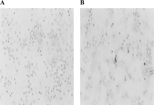
FIG. 1 Induction of apoptosis in White Leghorn lymphocytes treated with 10−5 M deltamethin in vitro. Photomicrographs (at X400) are of avian lymphocytes after immunoperoxidase staining to detect presence of Annexin-V (A) Control lymphocytes. (B) Lymphocytes treated with deltemethrin with translocated phosphatidylserine on their surface.

FIG. 2 DNA fagmentation in avian lymphocytes exposed to 10−5 M of deltamethrin. After treatments, DNA was harvested and 20 μg per sample electrophoresed over a 1% agarose gel. Lane 1: GeneRuler 100 bp DNA Ladder Plus Marker; Lane 2: DNA from control cells; Lane 3: DNA from cells after exposure to deltamethrin.
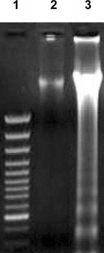
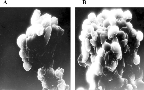
FIG. 2 DNA fagmentation in avian lymphocytes exposed to 10−5 M of deltamethrin. After treatments, DNA was harvested and 20 μg per sample electrophoresed over a 1% agarose gel. Lane 1: GeneRuler 100 bp DNA Ladder Plus Marker; Lane 2: DNA from control cells; Lane 3: DNA from cells after exposure to deltamethrin.
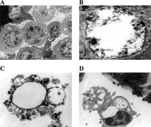
FIG. 3 Scanning electron microscopy (SEM) photographs showing apoptosis in avian lymphocytes induced by deltamethrin. (A) Budding of apoptotic bodies from lymphocyte (X15,000); (B) Apoptotic bodies on lymphocyte surface (X15,000).

FIG. 3 Scanning electron microscopy (SEM) photographs showing apoptosis in avian lymphocytes induced by deltamethrin. (A) Budding of apoptotic bodies from lymphocyte (X15,000); (B) Apoptotic bodies on lymphocyte surface (X15,000).

FIG. 4 Transmission electron microscopy (TEM) photographs showing apoptosis in avian lymphocytes induced by deltamethrin. (A) Control lymphocytes (X11,000). (B) Margination of chromatin (X15,000). (C) Formation of apototic bodies (X11,000). (D) Phagocytosis of apoptotic bodies (X11,000).

FIG. 4 Transmission electron microscopy (TEM) photographs showing apoptosis in avian lymphocytes induced by deltamethrin. (A) Control lymphocytes (X11,000). (B) Margination of chromatin (X15,000). (C) Formation of apototic bodies (X11,000). (D) Phagocytosis of apoptotic bodies (X11,000).

Reprints and Corporate Permissions

Please note: Selecting permissions does not provide access to the full text of the article, please see our help page How do I view content?

To request a reprint or corporate permissions for this article, please click on the relevant link below:

Academic Permissions

Please note: Selecting permissions does not provide access to the full text of the article, please see our help page How do I view content?

Obtain permissions instantly via Rightslink by clicking on the button below:

If you are unable to obtain permissions via Rightslink, please complete and submit this Permissions form. For more information, please visit our Permissions help page.