Figures & data
Fig. 1. Electrocardiogram at admission of the first case showing ST-segment elevation in the leads V1 and V2.
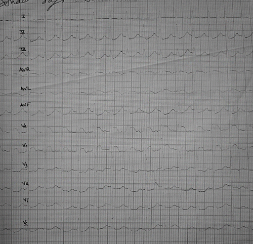
Fig. 2. Electrocardiogram of the first patient four hours after admission showed an increase of ST-segment in the leads V1 and V2.
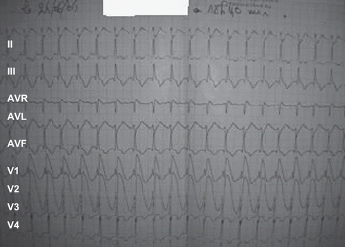
Fig. 3. Electrocardiogram in admission of the second patient showing ST-segment elevated in leads V1, V2, and V3.
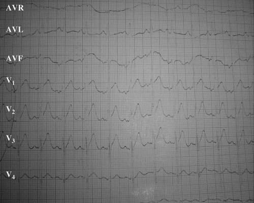
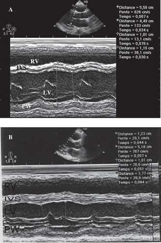
Fig. 4. Echocardiographically parasternal long axis depicted improvement of ventricle function in the second case. A) Echocardiogram obtained on the second day after admission depicts global hypokinesis with LVEF of 30% (calculated by Simpson method) and dilatation of LV. B) Echocardiogram taken eight days after admission showed improvement of ventricle function LVEF of 30%. RV: right ventricle; IVS: interventricular septum; LV: left ventricle; PW: posterior wall.