3,730
Views
10
CrossRef citations to date
0
Altmetric
Original Articles

Advanced Imaging and Robotics Technologies for Medical Applications

&
Pages 299-321 | Published online: 14 Dec 2011

Figures & data

Figure 1 Medical images; (a) US for liver phantom, (b) CT for ear, and (c) MRI for abdomen (color figure available online).
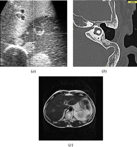
Figure 1 Medical images; (a) US for liver phantom, (b) CT for ear, and (c) MRI for abdomen (color figure available online).

Figure 2 Intra-operative MRI and surgical bed installed in the operating room (color figure available online).
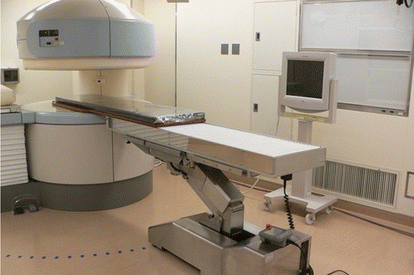
Figure 2 Intra-operative MRI and surgical bed installed in the operating room (color figure available online).

Figure 3 DTI image for brain surgery; nerve fibers are displayed (color figure available online).

Figure 3 DTI image for brain surgery; nerve fibers are displayed (color figure available online).

Figure 4 The surgical navigation; (a) configuration of image guidance for surgery, (b) display of surgical navigation for ear surgery (color figure available online).
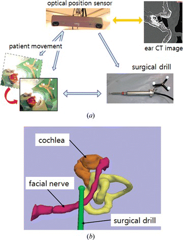
Figure 4 The surgical navigation; (a) configuration of image guidance for surgery, (b) display of surgical navigation for ear surgery (color figure available online).

Figure 5 Tool tracking system; (a) optical system vs. (b) electromagnetic system (color figure available online).
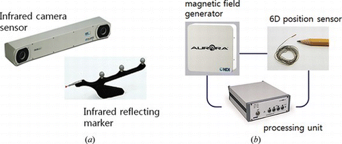
Figure 5 Tool tracking system; (a) optical system vs. (b) electromagnetic system (color figure available online).

Figure 6 Fiducial markers for registration; (a) skin markers, (b) template, and (c) anatomical landmarks on the temporal bone (color figure available online).
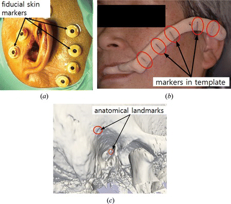
Figure 6 Fiducial markers for registration; (a) skin markers, (b) template, and (c) anatomical landmarks on the temporal bone (color figure available online).

Table 1. Advantages and drawbacks of registration methods

Figure 7 The display methods for image guided surgery; (a) multi-planar, (b) 3-D graphic, (c) augmented reality mode (color figure available online).
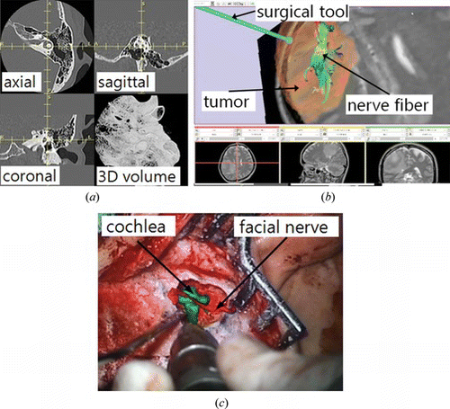
Figure 7 The display methods for image guided surgery; (a) multi-planar, (b) 3-D graphic, (c) augmented reality mode (color figure available online).

Figure 8 The first MRI compatible needle insertion robot (color figure available online).

Figure 8 The first MRI compatible needle insertion robot (color figure available online).

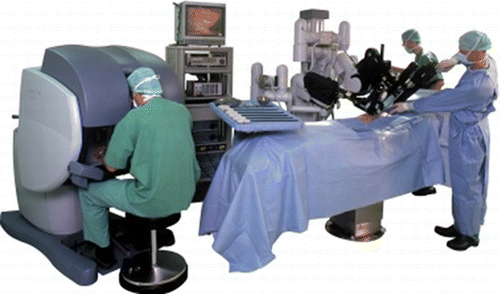
Figure 9 Master-slave surgical robot: daVinci (daVinci 2011) (color figure available online).

Figure 9 Master-slave surgical robot: daVinci (daVinci 2011) (color figure available online).

Figure 10 Forceps robot for endoscopic fetus surgery; small diameter, 2DOF, rigid forceps robot with LASER fiber (color figure available online).
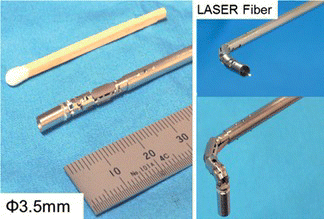
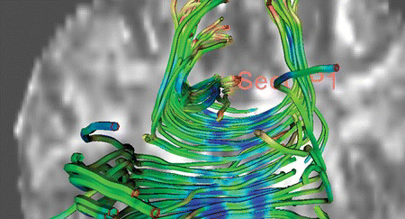
Figure 10 Forceps robot for endoscopic fetus surgery; small diameter, 2DOF, rigid forceps robot with LASER fiber (color figure available online).
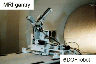
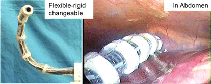
Figure 11 Flexible-rigid changeable endoscope guide for NOTES/SPS surgery (color figure available online).

Figure 11 Flexible-rigid changeable endoscope guide for NOTES/SPS surgery (color figure available online).

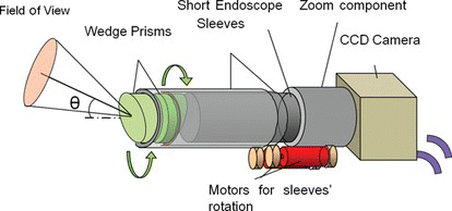
Figure 12 The concept of endoscope robot using two wedge prisms: By rotating two wedge prisms, arbitrary view is obtained without moving the endoscope itself (color figure available online).

Figure 12 The concept of endoscope robot using two wedge prisms: By rotating two wedge prisms, arbitrary view is obtained without moving the endoscope itself (color figure available online).

Reprints and Corporate Permissions

Please note: Selecting permissions does not provide access to the full text of the article, please see our help page How do I view content?

To request a reprint or corporate permissions for this article, please click on the relevant link below:

Academic Permissions

Please note: Selecting permissions does not provide access to the full text of the article, please see our help page How do I view content?

Obtain permissions instantly via Rightslink by clicking on the button below:

If you are unable to obtain permissions via Rightslink, please complete and submit this Permissions form. For more information, please visit our Permissions help page.