2,806
Views
55
CrossRef citations to date
0
Altmetric
Original Investigations

Schizophrenia: From the brain to peripheral markers. A consensus paper of the WFSBP task force on biological markers

, , , , , , , , , , , , , , , , , , , , & show all
Pages 127-155 | Published online: 12 Jul 2009

Figures & data

Figure 1.  The proposed cortical-subcortical circuitry implicated in schizophrenia. Relevant dopaminergic, glutamatergic, and GABAergic projections are depicted with bulky, fine, and dotted arrows, respectively. The nucleus accumbens, which receives extensive dopaminergic input from the ventral tegmental area (VTA) of the midbrain and prominent excitatory input from cortical areas, the prefrontal cortex (PFC), the hippocampus, and the amygdala, is thought to play a crucial role in integrating prefrontal and limbic input. Inhibitory projections from the nucleus accumbens are sent to the ventral pallidum, which projects to specific nuclei of the thalamus, which in turn send excitatory projections to various regions of the PFC. Dopaminergic projections from the VTA also innervate the PFC. Interactions between the PFC, the amygdala, and the hippocampus are not depicted (adopted from Thompson et al. Citation2004).

Figure 1.  The proposed cortical-subcortical circuitry implicated in schizophrenia. Relevant dopaminergic, glutamatergic, and GABAergic projections are depicted with bulky, fine, and dotted arrows, respectively. The nucleus accumbens, which receives extensive dopaminergic input from the ventral tegmental area (VTA) of the midbrain and prominent excitatory input from cortical areas, the prefrontal cortex (PFC), the hippocampus, and the amygdala, is thought to play a crucial role in integrating prefrontal and limbic input. Inhibitory projections from the nucleus accumbens are sent to the ventral pallidum, which projects to specific nuclei of the thalamus, which in turn send excitatory projections to various regions of the PFC. Dopaminergic projections from the VTA also innervate the PFC. Interactions between the PFC, the amygdala, and the hippocampus are not depicted (adopted from Thompson et al. Citation2004).

Table I.  Imaging studies of striatal presynaptic dopamine parameters in drug-naive (DN), drug free (DF) and treated (T) patients with schizophrenia.

Figure 2.  Schizophrenia-susceptibility genes and synaptic plasticity: The glutamatergic synapse with a hypothetical scenario in which genes (in italics) may have shared effects on synapses via influences on their formation, plasticity or signalling properties. Solid arrows point to direct interactions, dotted ones to indirect action. mGluR: metabotropic glutamate receptor, NMDA: ionotropic N-methyl-D-aspartate receptor, P5C: ▵1pyrroline-5- carboxylate; PRODH: proline dehydrogenase. (adopted from Harrison and Owen Citation2003).

Figure 2.  Schizophrenia-susceptibility genes and synaptic plasticity: The glutamatergic synapse with a hypothetical scenario in which genes (in italics) may have shared effects on synapses via influences on their formation, plasticity or signalling properties. Solid arrows point to direct interactions, dotted ones to indirect action. mGluR: metabotropic glutamate receptor, NMDA: ionotropic N-methyl-D-aspartate receptor, P5C: ▵1pyrroline-5- carboxylate; PRODH: proline dehydrogenase. (adopted from Harrison and Owen Citation2003).

Table II.  Glutamate parameters measured in brain tissue of schizophrenia patients

Figure 3.  The reelin cascade in the aetiopathology of schizophrenia. Reelin binds to the very-low-density lipoprotein receptor (VLDLR) and APOE receptor 2 (APOER2) that results in the activation of the cytoplasmic DAB1 (adapter protein disabled 1). Phosphorylated DAB1 activates phosphatidylinositide 3-kinase (PI3K) and subsequently AKT (V-akt murine thymoma viral oncogene homologue). AKT activation inhibits the activity of glycogen synthetase kinase 3 β (GSK3β) resulting the inhibition of t phosphorylation that promotes microtubule stability. DAB1 also activates SRC family tyrosine kinases (SFKs) following NMDAR phosphorylation which potentiates NMDAR-mediated Ca2+ influx. Elevated Ca2+ activates cAMP-response element binding protein (CREB) that initiates expression of important genes for synaptic plasticity, neurite growth, and dendritic spine development (adopted from Herz and Chen Citation2006).

Figure 3.  The reelin cascade in the aetiopathology of schizophrenia. Reelin binds to the very-low-density lipoprotein receptor (VLDLR) and APOE receptor 2 (APOER2) that results in the activation of the cytoplasmic DAB1 (adapter protein disabled 1). Phosphorylated DAB1 activates phosphatidylinositide 3-kinase (PI3K) and subsequently AKT (V-akt murine thymoma viral oncogene homologue). AKT activation inhibits the activity of glycogen synthetase kinase 3 β (GSK3β) resulting the inhibition of t phosphorylation that promotes microtubule stability. DAB1 also activates SRC family tyrosine kinases (SFKs) following NMDAR phosphorylation which potentiates NMDAR-mediated Ca2+ influx. Elevated Ca2+ activates cAMP-response element binding protein (CREB) that initiates expression of important genes for synaptic plasticity, neurite growth, and dendritic spine development (adopted from Herz and Chen Citation2006).

Table III.  Synopsis of selected characteristics of candidate endophenotypes in schizophrenia research (pers. comm. A. Jablensky)

Table IV.  CSF proteins and lipids involved in the pathophysiology of schizophrenia. Several CSF proteins and lipids are important in transport, nerve cell excitability, nerve cell growth and neuron secretion. These functions of lipids may have significant physiologic ramifications in schizophrenia (pers. comm. A. Fonteh)

Figure 4.  Chromosomal susceptibility loci for schizophrenia derived by meta-analyses of genomewide linkage scans, and proposed risk genes (pers. comm. M. Gawlik).

Figure 4.  Chromosomal susceptibility loci for schizophrenia derived by meta-analyses of genomewide linkage scans, and proposed risk genes (pers. comm. M. Gawlik).

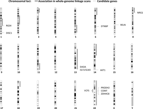
Table V.  Candidate genes for schizophrenic psychoses and strength of the biological evidence

Figure 5.  The potential role of cytokines in the pathophysiology of schizophrenia (pers. comm. YK Kim).

Figure 5.  The potential role of cytokines in the pathophysiology of schizophrenia (pers. comm. YK Kim).

Reprints and Corporate Permissions

Please note: Selecting permissions does not provide access to the full text of the article, please see our help page How do I view content?

To request a reprint or corporate permissions for this article, please click on the relevant link below:

Academic Permissions

Please note: Selecting permissions does not provide access to the full text of the article, please see our help page How do I view content?

Obtain permissions instantly via Rightslink by clicking on the button below:

If you are unable to obtain permissions via Rightslink, please complete and submit this Permissions form. For more information, please visit our Permissions help page.