Figures & data
Figure 1. The laboratory set for the exposure of the animals to phosphide.
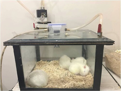
Table 1. Effect of aluminum phosphide (ALP) on cell number and cell size of adipocytes.
Table 2. Effect of aluminium phosphide on different parameters of biochemical in rats.
Figure 2. Effect of aluminum phosphide (ALP) on cell number and cell size of adipocytes of perirenal and perigonadal area.
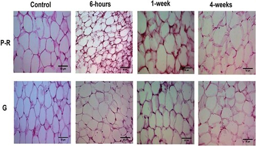
Figure 3. Liver tissue morphology in the rat in acute and sub-chronic period; (A): control group (×100) shows focal necrosis of the liver cells. The lobe of the vein is also seen in the lobular centre. Sinusoids are also dilated and there is no lupus necrosis, (B): One-week exposure (×400) severe necrosis and extensive necrosis of the liver are observed. The necrosis around the portal vein is probably due to the high amount of poison. Complex cells have increased in non-necrotic areas. Sinusoids are also disrupted. The lining of the vein of the lobular system is observed and cells have oedema, (C): 6h exposure (×100), (D): 4 weeks exposure (×100) damage to the liver cells is extensive and from the portal to the venous portal of the lobule centre, but is more intense around the vein of the lobular centre. Fatty change is also observed. Apoptosis and necrosis are observed remarkably in 10% of the liver cells, along with compensatory regenerative changes. The most important lesson of the necrosis of the bridge between the pleasures of the port, which indicates irreversible damage, leads to cirrhosis of the liver.
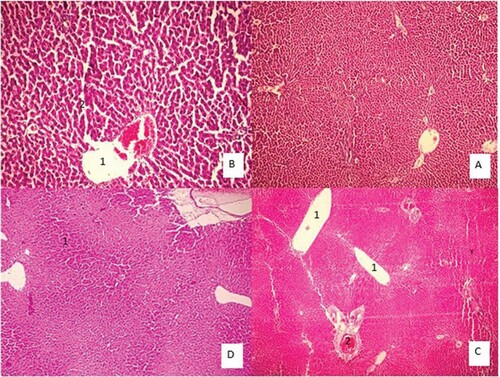
Figure 4. Lung tissue morphology in the rat in acute and sub-chronic period; (A): control group (×100), (B): One-week exposure (×400) shows alveolar walls are damaged. Contrary to the normal tissue, Type II pneumocystis increased and covers about 30% of the alveolar surface. Some alveoli are full of discharge and are seen in some types of bleeding, (C): 6h exposure (×100) shows Type II pneumocystis covers about 80% of the alveolar surface, resulting in a thickening of the alveoli. Spreading within the alveoli, severe vessel dilatation, and alveolar wall necrosis are also observed. Alveolar injury is more of an interstitial type that is in the vicinity of airways with greater intensity than near the pleural area, and (D): 4 weeks exposure (×40) shows a large number of interstitial (70% lung tissue) focuses on increasing intra-alveolar secretions. Pneumocystis Type II covers approximately 30% of the alveolar levels. Small haemorrhage can also be seen.
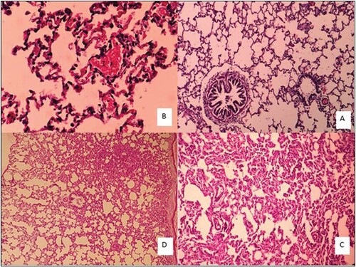
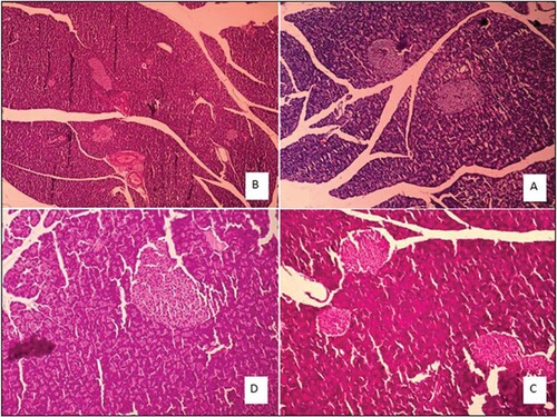
Figure 5. Pancreatic tissue morphology in the rat in an acute and sub-chronic period; (A): control group (×40), (B): One-week exposure, (C): 6h exposure, (D): 4 weeks exposure.