Figures & data
Figure 1. Crassostrea gigas D-shaped shell larvae (48 h post fertilization, hpf). Anatomy of the main organs constituting the digestive apparatus as visible through the clear shell by inverted light microscopy (Zeiss Axio Observer Inverted Microscope). (A) Oyster larva under protruded body position. (B) Oyster larva under retracted body position (position taken before death by all organisms). The black spot in the style sac area is a dense accumulation of ingested NPs. A: anus; E: esophagus; DG: digestive gland; I: intestine; M: mouth; MC: mantle cavity; S: stomach; SS: style sac; V: velum; VC: visceral cavity.
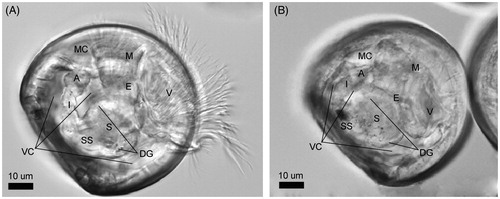
Figure 2. Ingestion of NPs and their internalization in the absorptive cells of the digestive gland. (A) Overview of the distribution of NPs inside the stomach lumen and the adjoined digestive gland. (B) Ingested NPs and bacteria in the proximity of the edge between the gastric shield and the style sac. (C) Entry of an ingested bacterium into an absorptive cell via phagocytosis. (D) Primary phagosome whose marked multi-lobe shape suggests recent fusion of pinosomes. (E) Progressive maturation of the endocytic bodies along their migration from the apical side to the basal side of an absorptive cell (i.e. pinosomes, early heterophagosomes, rounded-shape mature heterophagosomes). Pictures were taken from a grid stained with lead citrate. Arrows indicate NPs. ib: internalized bacterium; b: bacterium; m: mitochondrion; hp: heterophagosome; c: cilium; ld: lipid droplet; rer: rough endoplasmic reticulum.
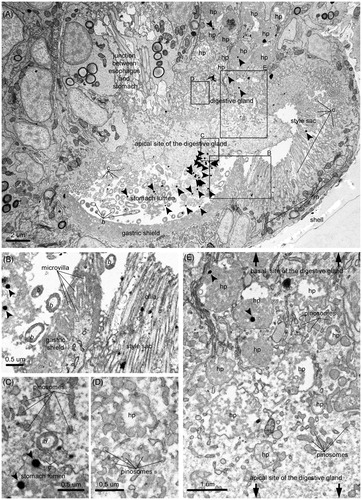
Figure 3. Intracellular digestion of NPs. (A) Electron micrograph showing a section of the digestive gland and a portion of the circular section of the densely ciliated esophagus. The digestive cells are sectioned at their mid-/basal- side, featuring an abundance of heterolysosomes with NPs inside the majority of them. The filling of these endocytic bodies with bacteria at different levels of decomposition and flocculent electron-dense material of heterogenous texture suggest stages in the digestive process, as exemplified by the detailed pictures B–E. More precisely, (B) Initial stage of intracellular decomposition, (C) Intermediate stage, (D) Late stage (E) Final stage, i.e. NP containing residual body storing indigestible material. Sections were imaged without prior lead citrate contrasting. Arrows point to NPs. e: endosome; ib: internalized bacterium; b: bacterium; m: mitochondrion; hp: heterophagosome; hl: heterolysosome; rer: rough endoplasmic reticulum; ld: lipid droplet.
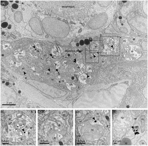
Figure 4. NP excretion. (A) NPs inside the lumen of the intestine and the residual bodies of epithelial cells. In the intestinal lumen, NPs are mixed with bacteria and the fecal laden mucous string. (B) Intestinal section containing bacteria, NPs, and feces. (C) NP containing residual bodies in the process of discarding their cargo in the lumen of the intestine. Pictures were taken from a grid stained with lead citrate. Arrows indicate NPs. hl: heterolysosome; b: bacterium; m: mitochondrion; rb: residual body; n: nucleus; c: cilium.
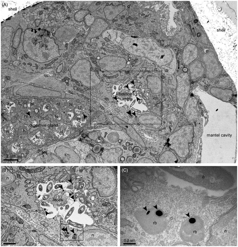
Figure 5. NP translocation. (A, C, E) NP containing residual bodies in cells near or lining the shell, identified as phagocytic coelomocytes. (B, D, F) Detailed images of the framed areas of pictures A, C, and E, respectively. Sections were imaged without prior lead citrate contrasting. Arrows point to NPs. m: mitochondrion; rb: residual body; n: nucleus; ld: lipid droplet; g: Golgi apparatus; rer: rough endoplasmic reticulum.
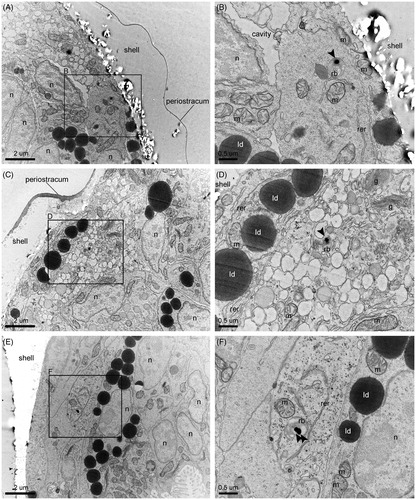
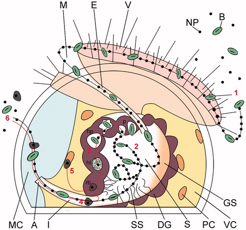
Figure 6. Schematic illustrating the proposed alimentary uptake pathway for NPs in Crassostrea gigas early veliger larvae. Sequential steps. (1) Capture and ingestion. Suspended particles are captured by the cilia of the velum, driven into the mouth and esophagus. (2) Post-ingestive sorting. Once inside the stomach, the ingested particles undergo vigorous swirling by the cilia of the style sac and post-ingestive sorting. Particles are directed toward the digestive gland for their cellular internalization, or toward the intestinal groove for excretion (see ). (3) Cellular uptake and intracellular digestion. Particles are taken up by the absorptive cells of the digestive gland via pinocytosis-macropinocytosis, gathered into heterophagosomes, and digested inside heterolysosomes; NPs, together with the residuals of the intradigestion process, are stored inside residual bodies (). (4) Clearance. NPs and non-nutritious material accumulated inside residual bodies are driven to the intestine for excretion with feces, along with non-absorbed particles sorted for excretion in the stomach (). (5) Translocation to phagocytic coelomocytes. Insoluble residuals of intracellular digestion, including NPs, are translocated to the phagocytic coelomocytes lining the inner membranes of the visceral cavity for processing or ejection via diapedesis (). (6) Excretion. Residuals of intradigestion and post-ingestive sorting are mixed into the fecal laden mucus string and ejected through the anus into the mantle cavity and eliminated from the larval body. A: anus; E: esophagus; DG: digestive gland; GS: gastric shield; I: intestine; M: mouth; MC: mantle cavity; PC: phagocytic coelomocyte; S: stomach; SS: style sac; V: velum; VC: visceral cavity; B: bacterium; NP: nanoparticle; p: pinocyte/macropinocyte; hp: heterophagosome; hl: heterolysosome; rb: residual body.