680
Views
0
CrossRef citations to date
0
Altmetric
Special Report

Recent insights into extracorporeal photopheresis for graft-versus-host disease

&
Pages 339-348 | Received 29 Sep 2023, Accepted 12 Dec 2023, Published online: 20 Feb 2024

Figures & data

Figure 1. ECP procedure and hypothesized mechanism of action. Step 1: Mononuclear cells (MNCs) are separated by peripheral blood by an apheresis cell separator. Step 2: MNCs are incubated with 8-methoxy psoralen that cross-links DNA bases after photoactivation by UV-A rays. The treatment induces cell apoptosis, maturation of tolerogenic dendritic cells (DC), and phagocytosis of apoptotic lymphocytes by DC. The process dampens inflammation by reducing pro-inflammatory cytokines, enhancing anti-inflammatory cytokines production, and promoting the expansion of Tregs which, in turn, produce anti-inflammatory cytokines. Step 3: treated cells are returned to the patient and 8-methoxypsoralen is inactiveFootnote1.

Figure 1. ECP procedure and hypothesized mechanism of action. Step 1: Mononuclear cells (MNCs) are separated by peripheral blood by an apheresis cell separator. Step 2: MNCs are incubated with 8-methoxy psoralen that cross-links DNA bases after photoactivation by UV-A rays. The treatment induces cell apoptosis, maturation of tolerogenic dendritic cells (DC), and phagocytosis of apoptotic lymphocytes by DC. The process dampens inflammation by reducing pro-inflammatory cytokines, enhancing anti-inflammatory cytokines production, and promoting the expansion of Tregs which, in turn, produce anti-inflammatory cytokines. Step 3: treated cells are returned to the patient and 8-methoxypsoralen is inactiveFootnote1.

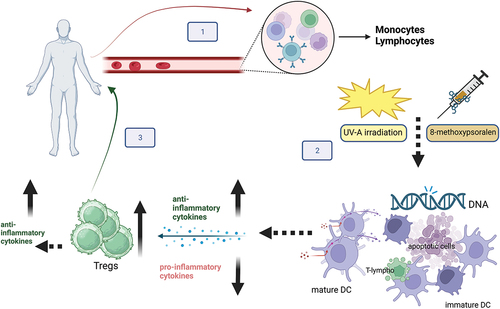
Table 1. Published studies on aGvhd and cGvhd patients treated by ECP.