Figures & data
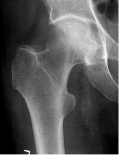
A conventional radiograph interpreted as being negative. In retrospect, it may be possible to see an irregularity in the proximal cortex of the femoral neck.
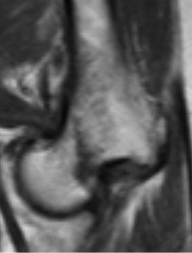
A T1 weighted image with a subcapital femoral neck fracture as a black line with a somewhat vertical progression through the femoral neck.
An STIR image showing high signal areas surrounding the fracture (white and light gray) and extending towards the lesser trochanter, indicating a recent trauma.
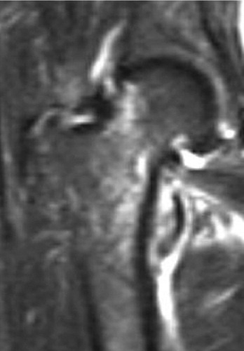
Table 1. Crosstable giving diagnoses after conventional radiography and the corresponding diagnoses after magnetic resonance imaging
Table 2. MRI diagnoses given by radiologist A and B (both experienced musculoskeletal radiologists) in 23 randomly selected examinations. Differences in interpretation are marked with red