Figures & data
Figure 1. Definition of ROI-1 and ROI-2 in radiographs and DXA scans.
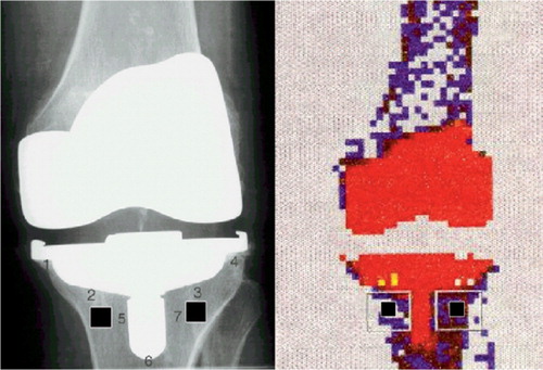
Figure 2. ROIs and density measurement by the optical method.
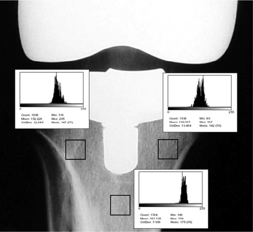
Table 1. Mean of density in the three ROIs at 12 and 24 months, and relationship regarding gender of patients
Table 2. Density obtained in the three ROIs, and average at 12 and 24 months
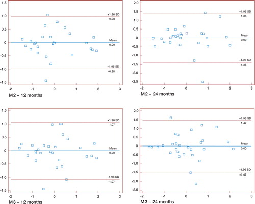
Figure 3. Bland and Altman plots for M2 and M3 at 12 and 24 months.Average of DXA (g/cm2) and optical density (gray-scale value) is shown on the horizontal axis.DXA (g/cm2) – optical density (gray-scale value) is shown on the vertical axis.