Figures & data
Fracture lines
Figure 1. The inverse T-fracture in 75° (A) and 150° (B) oblique view. Notice the ileum attached to the axial skeleton and with articular cartilage stretching into the joint posteriorly.
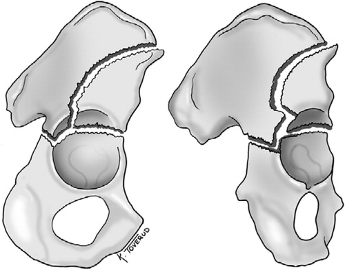
Figure 2. The transverse fracture with a floating acetabular dome in 75° (A) and 150° (B) oblique view. The acetabular dome is neither connected to the axial skeleton of the ileum nor to the ischium or the pubic bone.
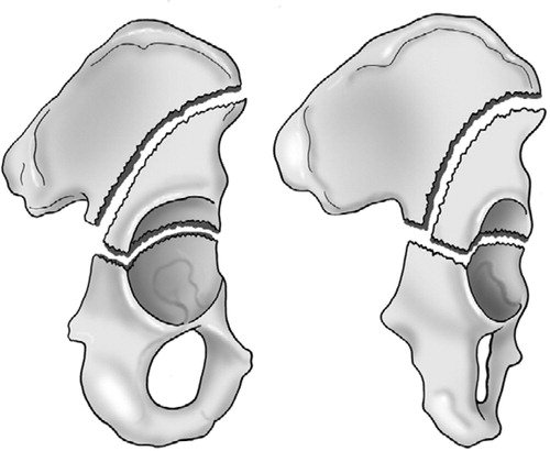
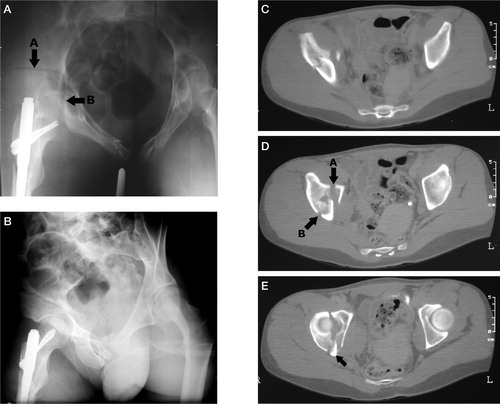
Figure 3. Patient no. 2 with an inverse T-fracture. In panel A, arrow A points to the vertical fracture line which divides and ascends to the crest and ant. sup. iliac spine. Arrow B points to the transverse fracture line. The axial scan in panel B shows the vertical fracture line which is also marked by arrow B in panel C. In this panel, arrow A marks the start of the transverse fracture. The posterior wall fragment is pointed out in panel D, and in panel E the distal end of the ileum with articular cartilage connected to the axial skeleton is marked with an arrow. Panel F shows the posterior wall fragment and there are no fracture lines involving the foramen obturatum.
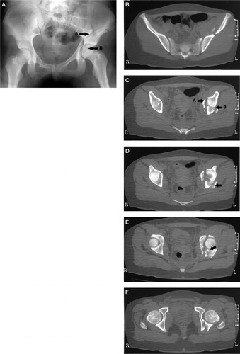
Figure 4. The inverse T-fracture of patient no. 4 is shown in panel A, and arrow A marks the vertical fracture line. Arrow B marks the transverse fracture line. On the oblique iliac view in panel B, the arrow points to the attached ileum with articular cartilage stretching into the joint posteriorly. In panel C, the vertical fracture is shown and both the vertical (A) and transverse (B) fracture lines are pointed out in panel D. The scan also shows comminution with an additional fragment marked C between the attached and unattached ileum. The posterior wall fragment is marked A in panel E and the arrow B points to attached ileum with articular cartilage. The most distal part of the attached ileum is marked in panel F. There are no fracture lines into the foramen obturatum.
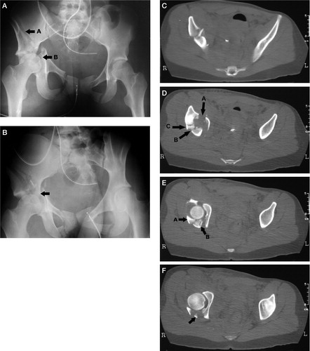
Figure 5. The transverse fracture with a floating acetabular dome is shown. In panel A with the pelvis inlet view, arrow A points to the vertical fracture line. Arrow B marks the transverse fracture line. (The inlet view is not part of standard radiographic examination of acetabular fractures.) In panel B, the iliac view clearly shows the vertical fracture line. The vertical line is also obvious on the axial CT-scan shown in panel C. Arrow A in panel D points to the start of the transverse fracture and arrow B shows the vertical fracture line. The scan is just proximal to the dome. In panel E, the arrow points to ileum connected to the IS-joint. No part of the acetabulum is attached to the axial skeleton.