Figures & data
Table 1. Clinical data before the first stage of reconstruction
Figure 1. Condition of soft tissue in case 4:chronic wound surrounded by scarred soft tissues on admission (A).Soft tissue defect after debridement and prior to coverage with a serratus anterior free flap.The cement spacer inlay has been implanted after a thorough bone debridement and stabilized with wires to the bony stumps.Tibial shaft stabilization is obtained with an external fixator (B).Complete skin graft healing over the free flap prior to removal of the cement spacer and bone reconstruction with iliac crest bone (C).
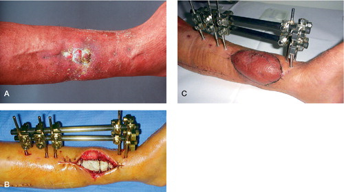
Figure 2. Case 4.Infected nonunion of the distal third of the tibia and nonunion of the fibula.The external fixator that was used initially had been removed before the patient was referred to us (A).After completion of the first-stage surgery (B).Bone healing at the last follow-up, 40 months after second-stage surgery (C).
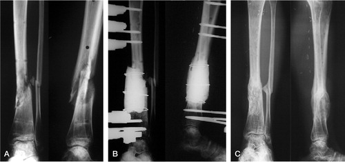
Table 2. Procedures and follow-up