Figures & data
Table 1. Patients at 6-year follow-up (FU) (n = 150) and 10-year follow-up (n = 118)
Table 2. Preoperative diagnoses (n = 118)
Figure 1. A. Illustration showing definition of radiolucent line (RL) and radiodense line (RD) around proximal stem. B. Illustration showing definition of osteolysis (longitudinal and focal) around proximal stem.
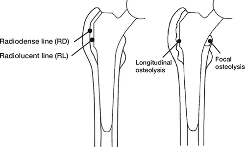
Figure 2. Radiolucent lines and osteolyses by Gruen zones. Comparison of results at 6–year and 10–year followup (FU).
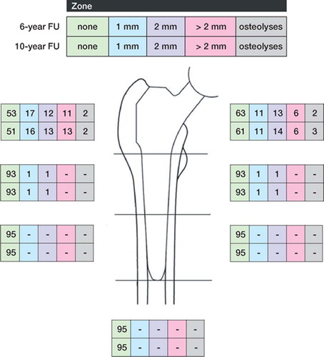
Figure 3. A) 50 year–old woman. Postoperatively. B) 1.5 years postoperatively. There was an RL of nearly 2 mm in zones 1 and 7. The boundary of the RL is a regular formed dense line (RD) corresponding to the preexisting bone stock, and running parallel to the implant surface. Atrophy of the medial cortical bone at zone 7. C) 4.5 years postoperatively. No change of RL and RD compared to B. At the distal end of the lines there is a small area of sclerosis. D) 10 years postoperatively. No change except a slight increase in the sclerosis in zone 7, thus reducing the extent of the RL and RD.
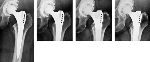
Table 3. Radiolucent lines and radiodense lines at 6-year and 10-year follow-up. Data in parentheses relate to radiolucent lines involving 50% or more of the zones affected (n = 95)
Figure 4. A) 65 year–old woman. Postoperatively. B) 3 years postoperatively: RL showing different diameters in zone 1; RL of 2 mm in zone 7. Inhomogeneous structure of RD in zone 1. C) 6 years postoperatively: increasing RL, in both width and distal extension, in zones 1 and 7. D) 10 years postoperatively: significantly increased dia meter of RL and widening also towards distally in zones 1 and 7. The corresponding RD showed an irregular density and distance to the implant surface.
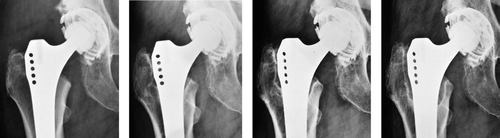
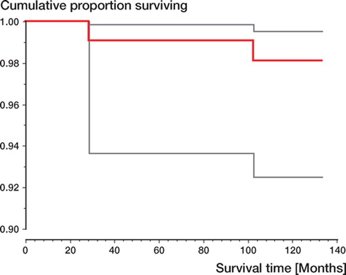
Figure 5. Kaplan-Meier survivorship curve showing 98% implant survival at 10 years with revision of the stem for any reason as the endpoint (95% CI: 99.5–92.5).