Figures & data
Figure 1. Typical microscopic appearance of the harvested periosteum. The periosteum contains two distinct layers: (A) a thick, outer fibrous layer, and (B) a thin, inner cambium layer which is adjacent to the bone (hematoxylin and eosin, × 200).
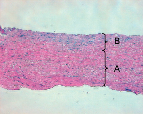
Figure 2. Typical macroscopic appearance of the regenerated tissue in the patellar grooves at 4 weeks (A) and 8 weeks (B) after surgery. A. The regenerated tissue appears slightly thicker than the surrounding normal articular cartilage.The regenerated cartilage-like tissue is opaque-white. B. The regenerated cartilage-like tissue appears translucent.The defect heals with smooth tissue, which is well integrated into the surrounding normal cartilage.
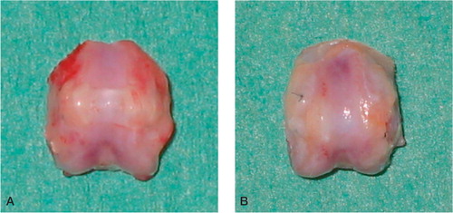
Figure 3. Typical light microscopic appearance (A, B) and fluorescent microscopic appearance (C, D) at 4 weeks after surgery.The arrow indicates the borderline between the repaired tissue and normal cartilage. The left side is repaired tissue. A. The defect is filled with the regenerated tissue that has been well stained by safranin-O.The regenerated cartilage-like tissue reaches over the level of the surrounding articular cartilage (safranin-O, × 100). B. The deep layer of the regenerated tissue has chondrocyte-like cells that are surrounded by a well-stained matrix, and the superficial layer of the regenerated tissue contains fibroblast-like cells.The regenerated cartilage-like tissue integrates well with the surrounding tissue (safranin-O, × 200). C. and D.Most cells in the regenerated tissue are GFP-positive cells derived from the donor periosteum in the superficial, middle, and deep region of the defect (C: × 100, D: × 200).
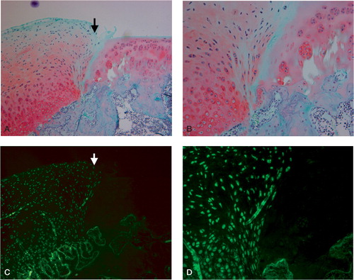
Figure 4. Typical light microscopic appearance (A, B) and fluorescent microscopic appearance (C, D) at 8 weeks after surgery.The arrow indicates the borderline between the repaired tissue and normal cartilage.The left side is repaired cartilage. A. The defect is repaired with regenerated cartilage-like tissue which integrates well with the surrounding normal cartilage (safranin-O, × 100). B. The deep layer of the regenerated tissue also has chondrocyte-like cells that are surrounded by a well-stained matrix (safranin-O, × 200). C. and D.Most cells in the regenerated tissue are GFP-positive cells derived from the donor periosteum in the superficial, middle, and deep region of the defect (C: × 100, D: × 200).
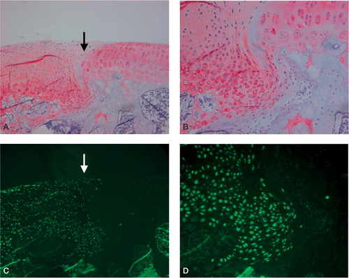
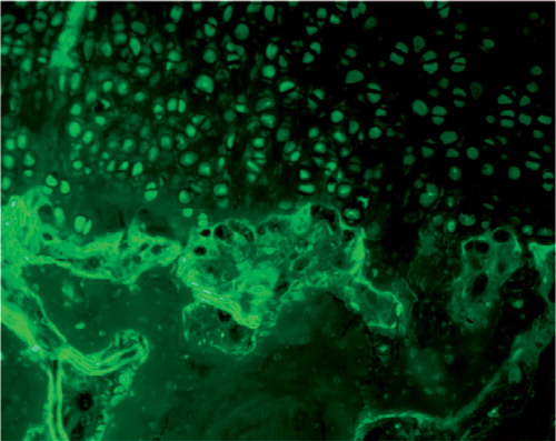
Figure 5. Fluorescent microscopic appearance of the border between regenerated tissue and the subchondral bone at 8 weeks after surgery. GFP-positive cells were not detected in the subchondral bone area below the regenerated tissue.