Figures & data
Figure 1. Anterior-posterior view and axillary view, immediately following closed reductio
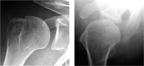
Figure 2. A and B: T1-weighted images; coronal view (A), axial view (B). C and D: T2-weighted images (soft tissue can be seen at tip of arrowhead); coronal view (C), axial view (D).
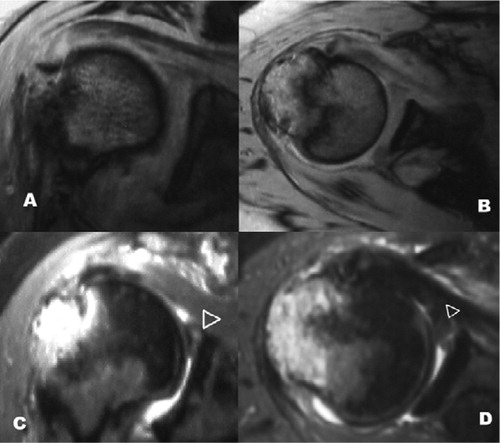
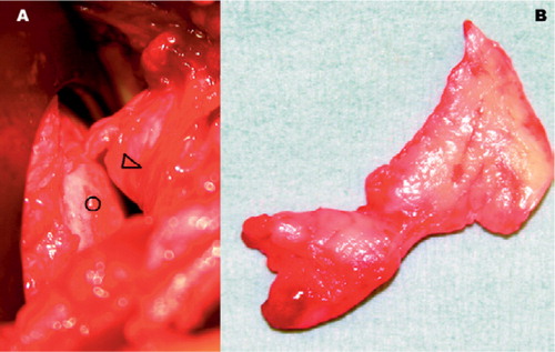
Figure 3. Intraoperative photographs. A: detached labrum (arrowhead) between humeral head (open circle) and glenoid, obscuring the glenoid surface. B: specimen of excised labrum.