Figures & data
Figure 1. A. Left humerus, AP view, at 2 months postoperatively (Oct 14,1986) showing good fit of the osteoarticular allograft into the glenoid fossa. B. 19 months postoperatively (May 9, 1988) showing subluxation of the humeral head. C. 15 years postoperatively (December 12, 2001). D. Specimen radiograph.A large osteophyte on the inferiormedial aspect of the humeral, subchondral sclerosis and areas of resorption are evident. E. Photomacrograph of a sagittal section of the explanted specimen showing remnants of soft tissue attachment to the graft.The bright yellow area (asterisk) corresponds to residual nonviable graft abutting the fibrovascular tissue (black arrow).The holes represent the sites of screw insertion for plate fixation. F. Low-power view of hematoxylin and eosin stained histological section of humeral head and proximal diaphysis showing nonviable graft (asterisk) separated from the viable areas by dense fibrovascular tissue (black arrow), which corresponds to the white region in E. An additional area of necrotic bone can be seen in the humeral head at the right.
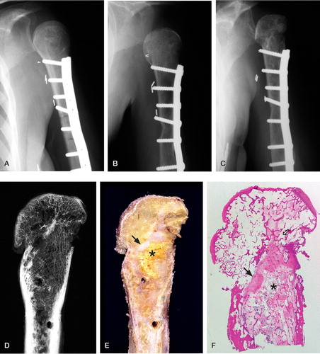
Figure 2. High-power (100 ×) view of fibrovascular tissue in marrow surrounding an allograft trabecula.The dense collagenous tissue is almost acellular, but contains numerous small blood vessel components.
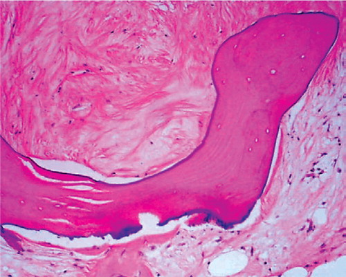
Figure 3. A. High-power (100×) view of the medial aspect of the proximal portion of the humeral head joint surface showing considerable eburnation.The subchondral bone is revascularized (black arrow) and contains numerous viable osteocytes. B. Low-power view depicting the viable osteophyte (black arrow). C. High-power (100×) of viable cortical and trabecular bone and mature bone marrow that is rich in vascular elements.
