Figures & data
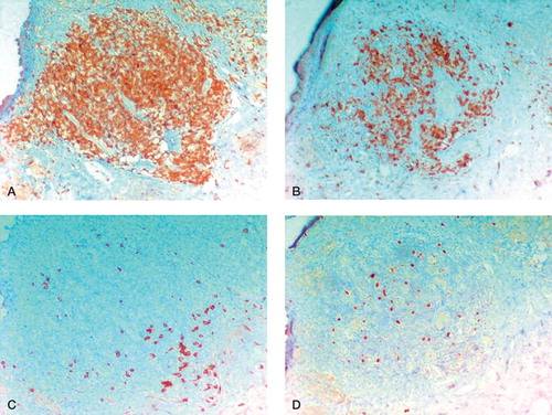
Immunohistochemical staining methods used for grading
Figure 1. Extent of necrosis in cases with in situ times of ≤ 12 months and < 12 months.
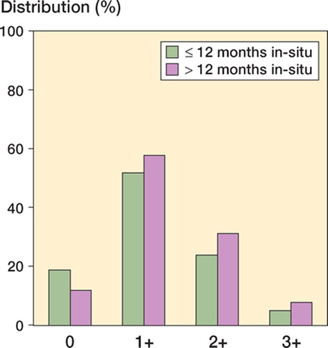
Figure 2. Storage of metallic particulate debris and histiocytic tissue reaction in cases with in situ times of ≤ 12 months and < 12 months.
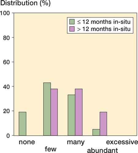
Figure 3. Micrographs of hematoxylin and eosin (H&E) stained sections (magnification 400×). A. Clusters of macrophages with “slate blue” cytoplasmic appearance. B. “Dusty” macrophage appearance, which is characterized by isolated black specks in the cytoplasm (white arrows). The particle size is up to 1 μm, while the normal size of lymphocytes is 5–7 μm (black arrows).
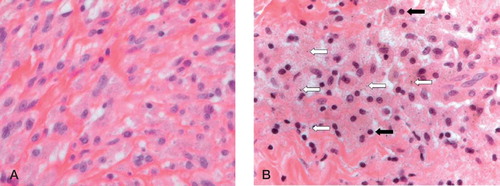
Figure 5. Perivasal accumulated lymphocytic infiltrates (H&E-stained sections). A. “Many” perivascular lymphocytic infiltrates (magnification 100×). B. “Abundant” perivascular lymphocytic infiltrates; few lymphocytic follicles without germinal centers (magnification 100×). C. and D. “Excessive” lymphoplasma cellular infiltration; lymphocytic follicles with formation of germinal centers in the intermediate vascular layer (magnification 25× (C) and 100× (D)).
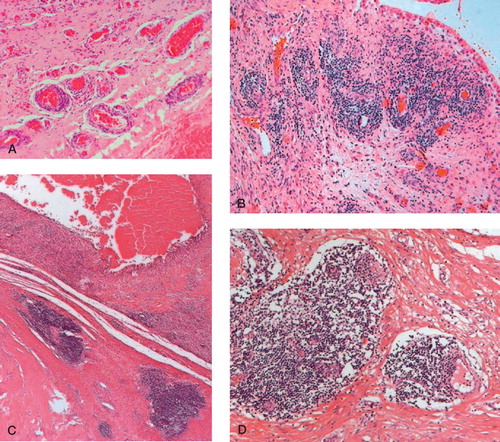
Figure 6. Different immunohistological stainings for a prominent synovial perivascular lymphatic follicle (magnification 100×). A. Follicular pattern of CD3-positive T-lymphocytes (brown-colored). B. Follicular pattern of CD20-positive B-lymphocytes. C. In the periphery, a small line of CD138-positive plasma cells. D. Increased number of MIB-1-positive lymphocytes showing proliferation.