2,000
Views
28
CrossRef citations to date
0
Altmetric
Original

Long-term results after closed reduction of latedetected hip dislocation: 60 patients followed up to skeletal maturity

&
Pages 236-246 | Received 20 Jun 2006, Accepted 12 Sep 2006, Published online: 08 Jul 2009

Figures & data

Figure 1. Radiographic measurements. Panels A and B show the primary radiograph of a 15-month-old girl with a dislocated right hip. AI is the acetabular index. The lateral (e) and vertical (f) metaphyseal distances are indicated.

Figure 1. Radiographic measurements. Panels A and B show the primary radiograph of a 15-month-old girl with a dislocated right hip. AI is the acetabular index. The lateral (e) and vertical (f) metaphyseal distances are indicated.

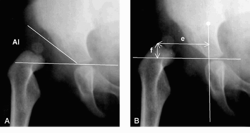
Figure 2. Radiograph of the same patient as in at the age of 9 years. Migration percentage (panel A) is a/b × 100 and indicates the percentage of the femoral epiphysis lateral to the lateral rim of the acetabulum. CE is center-edge angle and AA is acetabular angle (panel B)

Figure 2. Radiograph of the same patient as in Figure 1 at the age of 9 years. Migration percentage (panel A) is a/b × 100 and indicates the percentage of the femoral epiphysis lateral to the lateral rim of the acetabulum. CE is center-edge angle and AA is acetabular angle (panel B)

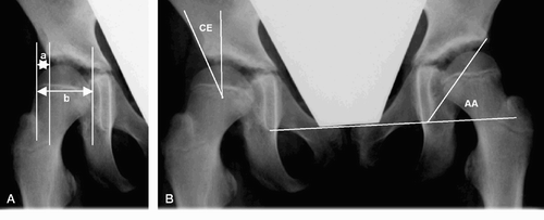
Table 1.  Radiographic measurements in 60 patients (78 abnormal hips and 42 normal hips) with late-detected hip dislocation. Results are given as mean (SD)

Table 2.  Radiographic results according to the Severin classification

Figure 3. Radiographs of a girl with a Spitzy shelf operation at the age of 12 years. Panel A shows acetabular dysplasia of the right hip before the shelf procedure was performed. Panel B, taken at the age of 17 years, indicates that the operation has given an adequate acetabular roof. Panel C shows no signs of osteoarthrosis at the age of 33 years.

Figure 3. Radiographs of a girl with a Spitzy shelf operation at the age of 12 years. Panel A shows acetabular dysplasia of the right hip before the shelf procedure was performed. Panel B, taken at the age of 17 years, indicates that the operation has given an adequate acetabular roof. Panel C shows no signs of osteoarthrosis at the age of 33 years.

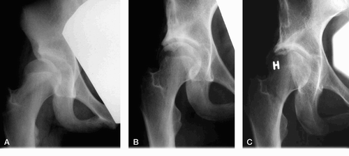
Table 3.  Radiographic parameters at the time of diagnosis and follow-up according to the outcome at skeletal maturity. Satisfactory outcome corresponds to Severin groups I–II; unsatisfactory outcome corresponds to Severin groups III–V. Results are given as mean (SD)

Reprints and Corporate Permissions

Please note: Selecting permissions does not provide access to the full text of the article, please see our help page How do I view content?

To request a reprint or corporate permissions for this article, please click on the relevant link below:

Academic Permissions

Please note: Selecting permissions does not provide access to the full text of the article, please see our help page How do I view content?

Obtain permissions instantly via Rightslink by clicking on the button below:

If you are unable to obtain permissions via Rightslink, please complete and submit this Permissions form. For more information, please visit our Permissions help page.