Figures & data
Figure 1. A. Dorsal buttress pin with washer and bicortical screw. B. Radial pin plate with pins and screws. C. Preoperative radiograph. D. The fixation device and the bone substitute in place.
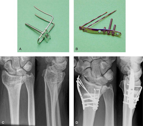
Figure 2 A. The operation was carried out with the arm in a prone position. 2 short incisions were made, one radially through the first extensor compartment and a second dorsal incision through the fourth compartment. B. The joint line was marked with 2 parallel pins into the distal radius and the holes were later used for the dorsal buttress pin. C. The osteotomy was then performed with an oscillating saw and the malposition reduced. A temporary fixation was achieved by a pin driven through the radial styloid and into the radius, proximal to the osteotomy, thereby bridging the osteotomy. D. When optimal positioning was accomplished, the fixation was secured by the dorsal buttress pin and the radial pin plate (E). F. Norian SRS was injected into the bone defect and left to harden as the extensor retinaculum and skin were closed. A bulky dressing and a dorsal cast completed the operation.
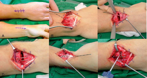
Table 1. Radiographic parameters preoperatively, postoperatively, and at final follow-up
Table 2. Objective results
Table 3. Subjective measures preoperatively, at 3 months, and at 1 year postoperatively