1,218
Views
28
CrossRef citations to date
0
Altmetric
Original paper

Sustained release of ciprofloxacin from an osteoconductive poly(DL)-lactide implant

, , , , , & show all
Pages 295-301 | Received 31 Mar 2007, Accepted 31 Mar 2007, Published online: 08 Jul 2009

Figures & data

Antimicrobial activity of ciprofloxacin released from the composite as a function of release time in vitro. The efficiency of unprocessed ciprofloxacin (control) is also shown. The values represent the lowest measured concentration (MIC) prohibiting bacterial growth of 5 standard ATCC strains

Figure 1. Release of ciprofloxacin from the pellets, expressed as a daily average release (μg/mL/day) and as a cumulative release (percentage of the total amount loaded in the pellets) as a function of in vitro immersion time in phosphate buffered solution.
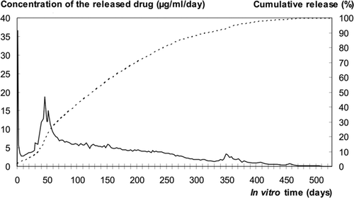
Figure 1. Release of ciprofloxacin from the pellets, expressed as a daily average release (μg/mL/day) and as a cumulative release (percentage of the total amount loaded in the pellets) as a function of in vitro immersion time in phosphate buffered solution.

Figure 2. SEM micrograph (100× magnification) of a composite microcylinder (pellet) at the start-up after melt-compounding. The PDLLA matrix mixed with bioactive glass microspheres showed microcavities and pores, which facilitated initial intrusion of fluid into the microstructure.
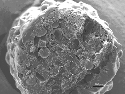
Figure 2. SEM micrograph (100× magnification) of a composite microcylinder (pellet) at the start-up after melt-compounding. The PDLLA matrix mixed with bioactive glass microspheres showed microcavities and pores, which facilitated initial intrusion of fluid into the microstructure.

Figure 3. SEM micrographs (500× magnification) of composite microcylinders as a function of immersion time in phosphate buffer. Consecutive representative specimens from two time points: immediately after processing (A) and at day 49 of the immersion test (B). The cut surfaces of the implants, examined under SEM, showed that the PDLLA matrix developed microporosity during degradation and release of ciprofloxacin.
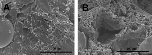
Figure 3. SEM micrographs (500× magnification) of composite microcylinders as a function of immersion time in phosphate buffer. Consecutive representative specimens from two time points: immediately after processing (A) and at day 49 of the immersion test (B). The cut surfaces of the implants, examined under SEM, showed that the PDLLA matrix developed microporosity during degradation and release of ciprofloxacin.

Reprints and Corporate Permissions

Please note: Selecting permissions does not provide access to the full text of the article, please see our help page How do I view content?

To request a reprint or corporate permissions for this article, please click on the relevant link below:

Academic Permissions

Please note: Selecting permissions does not provide access to the full text of the article, please see our help page How do I view content?

Obtain permissions instantly via Rightslink by clicking on the button below:

If you are unable to obtain permissions via Rightslink, please complete and submit this Permissions form. For more information, please visit our Permissions help page.