Figures & data
Figure 1. Low-power (A) and high-power (B) photomicrographs showing LYVE-1+ endothelial cells lining lymphatic vessels in the arthroplasty pseudocapsule. (Immunoperoxidase; 100× and 200× magnification).
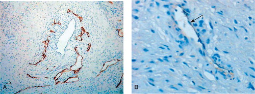
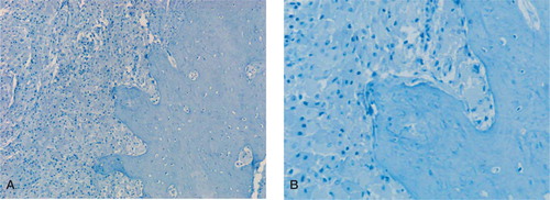
Figure 2. Low-power (A) and high-power (B) photomicrographs showing absence of LYVE-1 staining in the femoral arthroplasty membrane, which contains numerous plump (wear particle-containing) macrophages and surrounding compact cortical bone. (Immunoperoxidase; 100× and 200× magnification).