Figures & data
Figure 1. A and B. Schematic drawing and image representing anterior rabbit scapula with musculature and humerus removed demonstrating the bony tunnel of the subscapularis tendon. C. Demonstrates the rabbit scapula on end with an additional view of the bony tunnel. D and E. Schematic drawing and image representing the anterior rabbit scapula with the attached musculature.
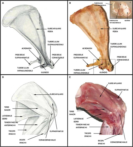
Figure 2. The top images represent the 3 phases of the right forelimb gait cycle in the rabbit, from paw strike to lift-off. Using video analysis, the scapulohumeral angle was calculated at each phase of the gait cycle. These angles were then translated to the dissected specimen (represented by the blue line on the rabbit forelimb). The lower series of images relates the translated scapulohumeral angle to the dissected specimen. The yellow dot represents the coracoid process and the black line the subscapularis tendon insertion on the humerus. Gross inspection reveals that the tendon has excursion within the tunnel from paw strike to lift-off. This is represented by the increase in distance from the yellow dot (corocoid process) to the black line (subscapularis insertion).
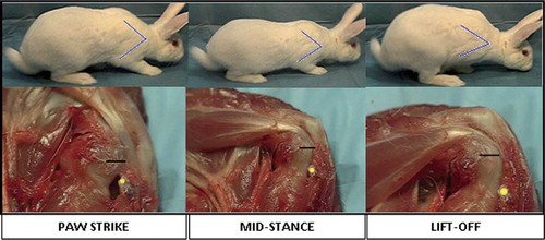
Figure 3. A. Anterior/posterior view subscapularis tendon insertion on proximal humerus. B. Humerus potted in polyvinylchloride pipe, substance of tendon sutured and secured with small frag plate. C. Instron machine. D. Humerus in position with tendon in clamp of Instron machine.
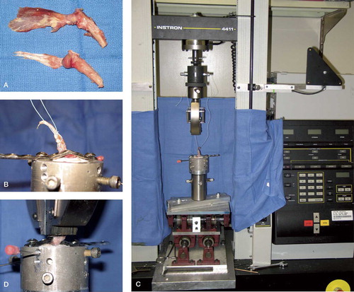
Figure 4. Force (N) versus displacement (mm) curve of rabbit subscapularis tendon. The average ultimate load was 112 N (SEM 23). The averaged ultimate deformation was 1.3 mm (SEM 0.15) and the stiffness (slope) was therefore 146 N/mm
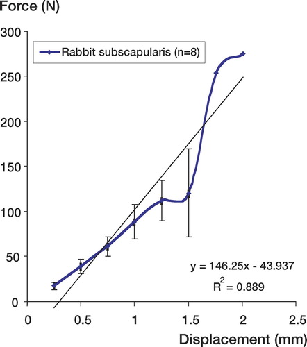
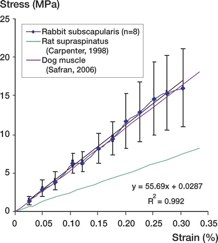
Figure 5. Stress (MPa) versus strain (%) comparing elastic modulus of rabbit subscapularis tendon to previous animal models for rotator cuff pathology. The rabbit subscapularis tendon has an elastic modulus of 56 MPa (R2=0.99). This modulus most closely correlates with the dog model for rotator cuff pathology 55 MPa.