Figures & data
Figure 1. Graphic representation of the retina layers with sub retinal fluid in CSC context.
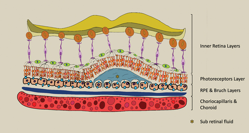
Figure 2. Optical coherence tomography of subretinal fluid in foveal zone. Pachychoroid is present.
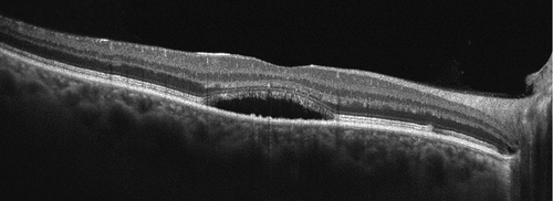
Figure 3. OCT representation of PED in parafoveal zone.
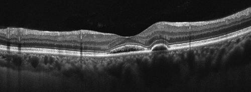
Figure 4. ICGA image showing hypecyanescient spot representing a leaking point in CSC patient.
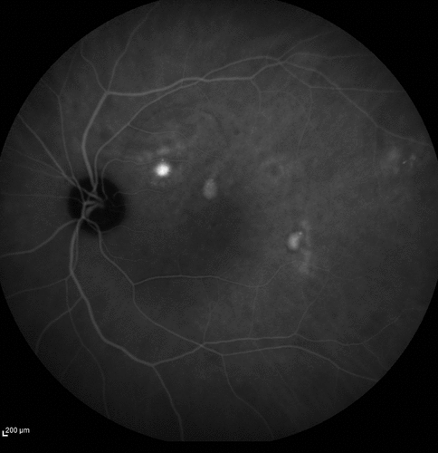
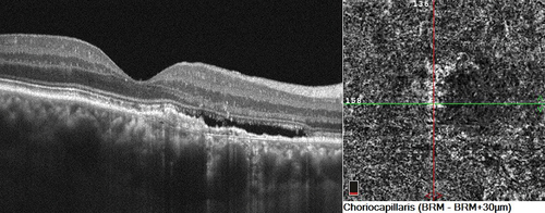
Figure 5. OCT representing a chronic CSC with slightly elevated RPE and SRF, the en face image of choriocapillaris enhance flow signal consistent with CVN.