Figures & data
Table 1.
Figure 1. (a) Case 1. Caudal right anterior oblique (RAO) coronary angiograms obtained in 2005 and 2015 revealing the appearance of a severe Medina 1,1,1 LMCA lesion. Result after provisional stenting. (b) Case 1. A triple X-kiss pattern is discovered at the balloons, Wires and Guiding catheters levels. Balloons and wires X-kissing was unexpected.
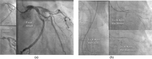
Figure 2. (a) Case 2. Caudal RAO and cranial LAO views before (a, b) and after stenting (c,d) a severe Medina 1,1,1 LMCA lesion, CX artery is a small-sized vessel. (b) Case 2. The triple X-kiss pattern (GCs, Wires and balloons) is again observed.
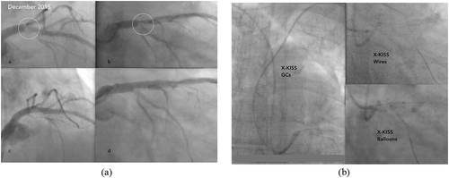
Figure 3. (a) Case 3. Cranial and caudal RAO views of a severe Medina 1,1,1 LMCA lesion before (a, b) and after T stenting (c, d). Early emergence of the first OM branch from the proximal CX artery and the large size of the vessels can be observed. View of the final X-kiss balloon inflation after T stenting (e). (b) Case 3. Triple X-kiss pattern again present. Triple wiring (LAD artery, CX artery, and first OM branch) was used for the trifurcated distal LMCA lesion.
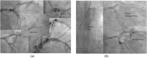
Figure 4. Case 4. Caudal RAO views before and after LAD/CX artery stenting. Final kiss balloons view (“V-Pattern”), only the GCs X-kissed.
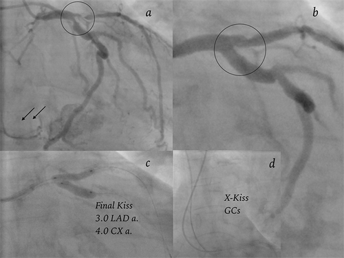
Figure 5. Case 5. Cranial RAO (40–10°) view of LCA, before and after distal, proximal and mid LAD artery stenting (P O T for LAD/Diag 2 and LAD/Diag 1 arteries).
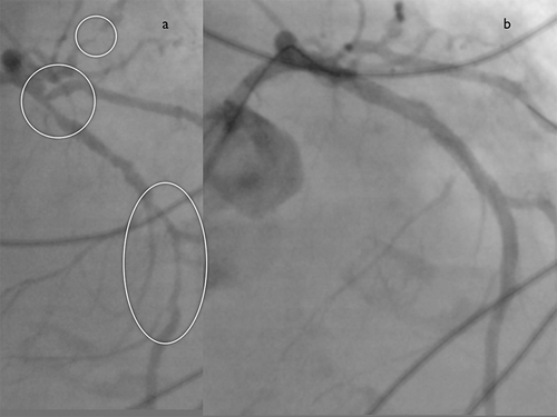
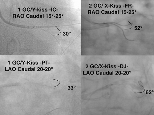
Figure 6. X versus Y-kiss at the wires level: 1 Guiding Catheter versus 2 Guiding Catheters in LAO and RAO caudal views. Y-kiss wires views are extracted from records of previous single access TRA cases of distal LMCA PCI.