Figures & data
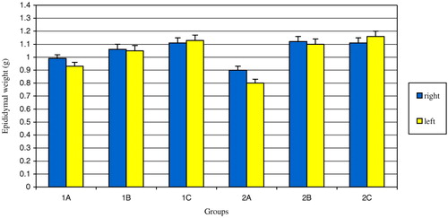
FIGURE 1 The comparison of the mean epididymal weights of the groups. The difference between right epididymal weights of groups 1A, 1B and 1C was statistically significant (p=0.007). The difference between left epididymal weights of groups 1A, 1B and 1C was statistically significant (p=0.006). The difference between right epididymal weights of groups 2A, 2B and 2C was statistically significant (p=0.009). The difference between left epididymal weights of groups 2A, 2B and 2C was statistically significant (p=0.003). The difference between right and left epididymal weights of group 2A was statistically significant (p < 0.05).
The Comparison of the Mean Tubular Diameters of the Epididymides (μm)
FIGURE 2 Apoptotic epididymal principal cell stained with TUNEL method. Black arrow shows apoptotic principal cell. Magnification (×400).
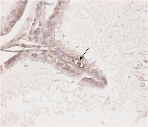
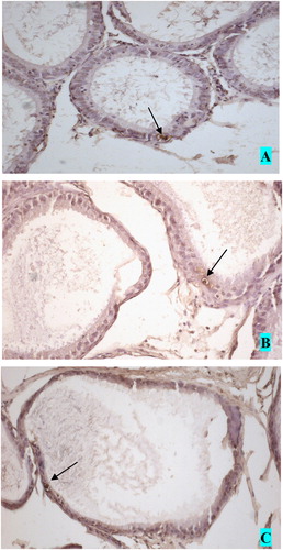
FIGURE 3 Apoptotic cells in the epididymal epithelium. A. caput, B. corpus and C. cauda epididymis. Magnification (×200).
TUNEL-Positive Cell Distribution in the Epididymal Epithelium. [0 is (−), 0.1–0.5 is (+), 0.51–1.0 is (++) and 1.0 < is (+++)]