Figures & data
Figure 1. Schematic representations of gap junction and connexins. Left: Gap junction between two cells. Each gap junction is composed of two connexons contributed by each cell. Individual connexon is made up of six protein subunits called connexins. Top-right: Structure of a connexin, which is a transmembrane protein with four transmembrane domains (TM1 to TM4). They are connected by two extracellular loops (EL-1 and EL-2) and one cytoplasmic loop (CL). Each connexin contains amino (NT) and carboxyl (CT) terminus in the cytoplasm. Bottom-right: ‘End-on’ view of a gap junction hemichannel (connexon). Two connexons align end-to-end to form a complete channel between neighboring cells. Each connexon is formed from six connexins.

Figure 2. 3D structure of Cx26 corresponding to a hemichannel, which is obtained from protein data bank (accession no. 2Zw3). Colors: red, O; white, H; cyan, C; blue, N; and yellow, S.
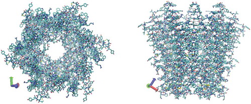
Figure 3. Atomic-scale representation of N-terminal of connexin Cx26, consisting of 20 amino acids. Its amino acid sequence is MDWGTLQTILGGVNKHSTSI.
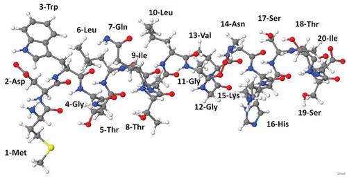
Figure 4. Snapshots from reactive MD simulations, presenting the breaking of a C-N bond in the backbone of N-terminal upon impact of an radical on the OH of Ser. (a) One
radical approaches OH residue in Ser. (b) H2O and O2 molecules are formed. (c) C-N bond is breaking and C-C double bond is forming. Colors: red, O; white, H; cyan, C; and blue, N.
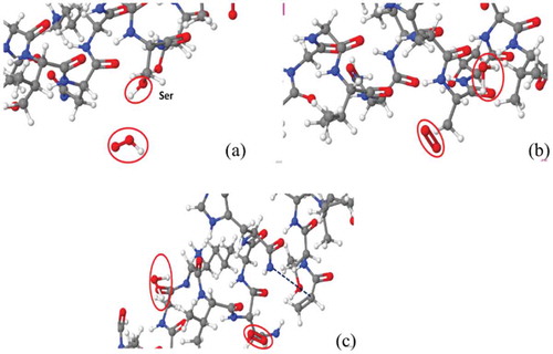
Figure 5. Reaction mechanism of C-N bond breaking upon impact of an radical on OH group in Ser, with the creation of the double C-C bond and the formation of a resonance-stabilized amide radical.
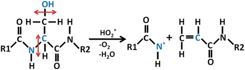
Figure 6. Snapshots from reactive MD simulations, presenting the breaking of a C-C bond in the backbone of N-terminal upon impact of three radicals on the hydrogen atom of Gly. (a) a carbonyl group is formed in Gly after the hydroxylated alpha-carbon reacts with a
radical. (b) C-C bond is breaking due to oxidation on Gly. Colors: red, O; white, H; cyan, C; and blue, N.
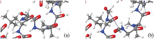
Figure 7. Reaction mechanism of C-C bond breaking upon three subsequent impact of *OH radical on the hydrogen atom in Gly.
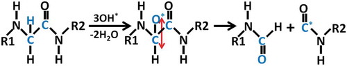
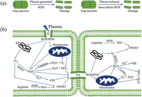
Figure 8. Plasma-generated ROS induced bystander effect via gap junction: (a) plasma-generated ROS and plasma-induced intracellular ROS resulting into the damage of gap junction; (b) cell death induced by plasma-oxidative stress in neighboring cells in mediated by a bystander effect. In the left cell, plasma-generated ROS stimulation of the mitochondria and endoplasmic reticulum (ER) leads to the generation of ROS. The transfer of ROS and RNS to neighboring cells via dysfunction GJ. Here, we show that plasma activated cell-cell communication between neighboring cells via dysfunction GJ.