Figures & data
Table 1. List of bacterial isolates and plasmids used in the G. mellonella infection assays.
Figure 1. Shigella spp. infection of G. mellonella induces strain dependent lethality. Panel A, Log phase cultures of S. flexneri 2a strain 2457T (––▪––), S. sonnei strain Moseley (–▴–), and S. dysenteriae 1 strain 1617 (•–▾–•) were harvested and 7 × 10⁵ CFU bacteria were injected into the larva. Each group contained 10 larvae. PBS mock injection was used as a negative control (--•--). Survival was monitored over 54 hours p.i. Panel B, photographic images of the G. mellonella larvae injected with 7×10⁵ CFU/larvae of 2457T, BS103 or PBS at 24 hours p.i., showing melanization of the larvae by injection of 2457T.
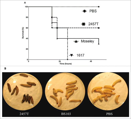
Figure 2. Dose, time and IpaB dependence of G. mellonella larvae death after infection with Shigella spp. G. mellonella larvae were injected with different concentrations of panel A, 2457T, panel B, BS103, panel C, 2457TΔipaB and panel D, 2457T ΔipaB : : ipaB. Larval death was monitored over time and scored by lack of movement after a touch. Significance was determined by the Log Rank test.
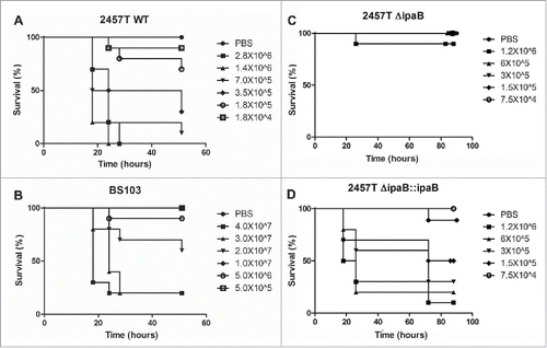
Table 2. LD50 of Shigella strains in the wax worm.
Figure 3. G. mellonella larval death is dependent on bacterial growth temperature and bacterial growth phase. G. mellonella larvae were injected with log (LP) and stationary phase (SP) 2457T that were grown at 37°C and 30°C. After injection, the larvae were incubated at either 37°C or 30°C, and death was monitored over time.
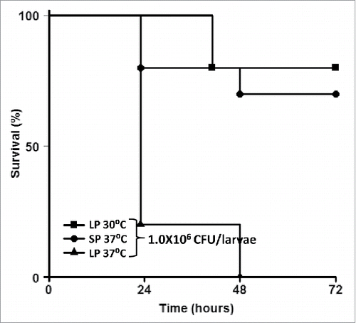
Figure 4. Bacterial replication and haemocyte depletion after Shigella infection in Galleria larva. G. mellonella larvae were infected with PBS, 1.0 × 10⁶ CFU of 2457T and 1.0 × 10⁶ CFU of BS103. After 24 hours, larvae survival rate, bacterial load in larvae and haemocyte counts were recorded. 4 live larvae at t = 0 and t = 24 were killed for these assays. Panel A represents percent survival of larvae at t = 0 and t = 24. Panel B represents changes in bacterial load in whole body at the 2 time points. Panel C represents per cent haemocyte counts at the 2 time points. These experiments were repeated 4 times. Each error bar represents one standard deviation from the mean. Differences between wild-type and BS103 were assessed for statistical significance by using a 2-sided t-test and assuming unequal variances. *p<0.05. Dark-filled bars t = 0, light gray bars, t = 24.
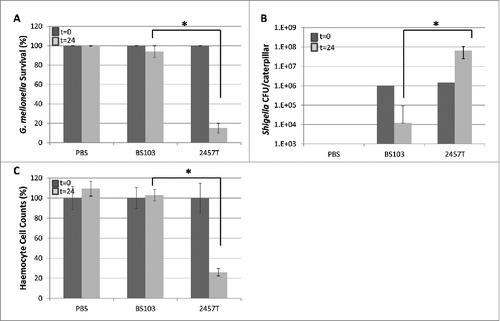
Figure 5. S. flexneri replicates within G. mellonella haemocytes. 2457T was tagged with a plasmid encoding GFP and used to infect larvae of G. mellonella at a dose of 1 × 10⁶ CFU/larvae. The larvae were killed 2 hours post infection. Samples of the larval hemolymph were observed using panel A, bright field and panel B, fluorescent microscopy. The merged image in panel C shows GFP-tagged bacteria within the haemocytes. Panels D, E, and F represent a different field where multiple haemocytes can be seen with GFP-laden bacteria. Panel F represents the merged image of panels D and E.
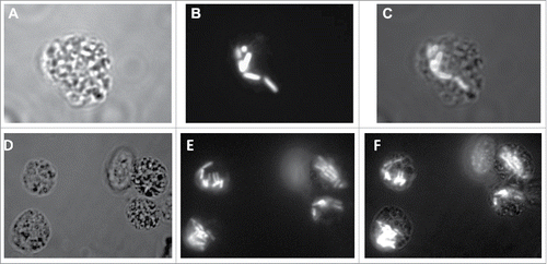
Figure 6. H&E-stained sections of G. mellonella after Shigella infection. G. mellonella were infected with 1 × 10⁶ CFU/larvae of 2457T. After 2 hours and 20 hours of growth at 37°C the larva were fixed and stained. Panel A, 2 hours p.i. Haemocytes slightly more clustered than observed in uninfected hemolymph (panel D). Panels B and C, 20 hours p.i. Haemocytes form nodules with evidence of melanization (black arrow in panel B and inset in panel B) and a load of bacteria around tubular organelle (red arrows in panel B). Panel D, PBS injected sample. No bacteria are observed in haemocoel and no obvious aggregates of haemocytes or melanization in uninfected samples.
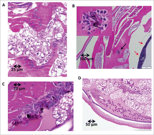
Figure 7. Immunofluorescence microscopy of 2457T-infected G. mellonella. G. mellonella larvae were infected with 1.0 × 10⁶ CFU of 2457T/larvae. After 20 hours the larvae were fixed and stained using monoclonal anti-S. flexneri LPS antibody as primary antibody and Alexa-fluor 594 conjugated secondary antibody. Panel A, autofluorescence of uninfected G. mellonella section, panel B, autofluorescence of infected larvae section, panel C, immunofluorescence of anti S. flexneri specific antibodies and panel D, higher-magnification view of the square marked region in panel C showing red-labeled bacteria clustering around tubular organs.
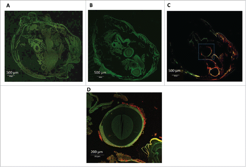
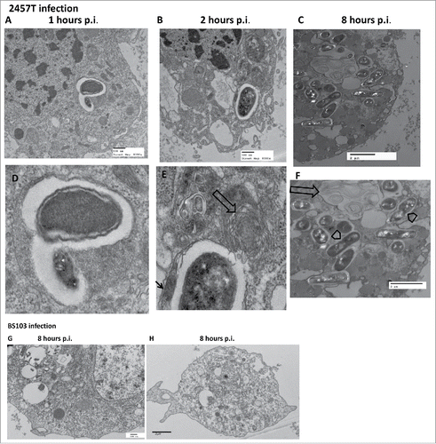
Figure 8. TEM of G. mellonella larval haemocytes after Shigella infection. Haemocytes of G. mellonella infected with 1.0 × 10⁶ CFU/larva, were fixed, sectioned and observed under the TEM. Panel A-F, sections of G. mellonella larval haemocytes infected with S. flexneri 2a 2457T. The lower panels D-F represent higher magnification images of bacteria-containing regions in the corresponding upper panels A-C (wide arrows-structures appearing as onion rings, arrow heads-replicating bacteria, thin arrow-fibrillar edges). Panels G-H, larval haemocytes infected with BS103.