
Figures & data
Figure 1. Illustration of superficial peroneal nerve anatomy and its branches, medial dorsal cutaneous and intermediate dorsal cutaneous nerves.
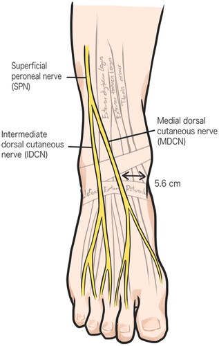
Figure 2. Illustration of SPNC flap designed and drawing. Skin flap was designed over the dorsalis pedis artery and the pedicle of flap was designed along the MDCN trajectory with 3 cm width (dotted line).
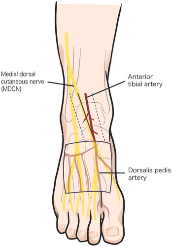
Figure 3. lateral side view of right foot shows colorized perforating branches of the dorsalis pedis artery under the pedicle of the flap (white asterisk). SPN, superficial peroneal nerve; P, pedicle of flap.
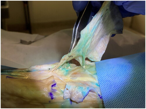
Figure 4. Exploration of superficial peroneal nerve underneath the flap shows the staining of paraneural vessels (asterisk) along the nerve and the neurocutaneous perforator (arrow) linking paraneural vessels and subcutaneous plexus.
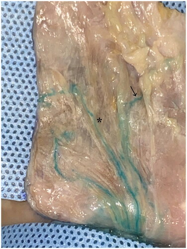
Figure 5. Illustration of the superficial peroneal neurocutaneous flap harvesting in proximally based design. SPN, superficial peroneal nerve; DPA, dorsalis pedis artery; F, skin flap; Asterisk (**), perforating branches from DPA and ATA.
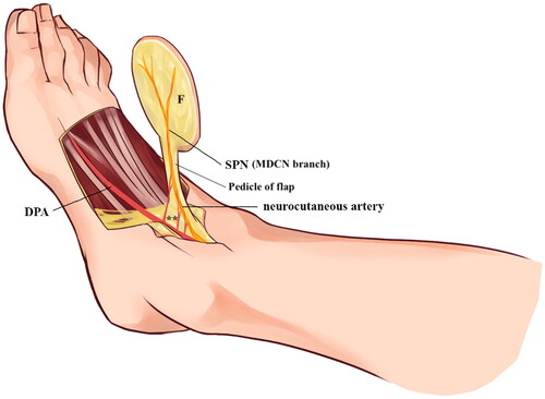
Table 1. cadaveric study results.