1,280
Views
1
CrossRef citations to date
0
Altmetric
Research Article

Ultrastructure of the dorsal claustrum in cat. I. Types of neurons

, , , , , ORCID Icon, , ORCID Icon & show all
Article: 1578636 | Received 21 Dec 2018, Accepted 30 Jan 2019, Published online: 27 Feb 2019

Figures & data

Fig. 1. An overview of the dorsal claustrum in cat. (a) A section through the brain showing the site (black arrow) from which the material for electron microscopy was obtained. (b) Histological control image of a Nissl stain showing the localization of the dorsal claustrum. Scale bar −500 µm.
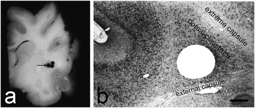
Fig. 1. An overview of the dorsal claustrum in cat. (a) A section through the brain showing the site (black arrow) from which the material for electron microscopy was obtained. (b) Histological control image of a Nissl stain showing the localization of the dorsal claustrum. Scale bar −500 µm.

Fig. 2. Multipolar and fusiform large neurons in the dorsal claustrum in cat. (a) Multipolar large neuron with an oval nucleus (N). n – nucleolus; D – dendrite; black arrowhead – glial cell. Scale bar − 3 μm. (b) Multipolar large neuron with an oval nucleus, electron-lucent nucleus (N). D – dendrites. Scale bar − 5.5 μm. (c) Multipolar large neuron with a typical Nissl body. Long black arrows – axo-somatic synaptic contacts; short black arrow – Golgi complex; black arrowhead – lysosomes. Scale bar − 2 μm. (d) Multipolar large neuron with prominent Golgi complex comprising cisterns and small electron-lucent vesicles (black arrows). Scale bar − 1.4 μm. (e) Fusiform large neuron with an oval nucleus (N) and an electron-dense nucleolus (n) containing fibrillar centers. The nuclear membrane forms invaginations of various sizes (long black arrows). Scale bar − 5.5 μm. (f) Fusiform large neuron with a slightly elongated nucleus (N). Long black arrows – invaginations of the nuclear membrane; short black arrows – axo-somatic synaptic contacts. Scale bar − 5.5 μm.
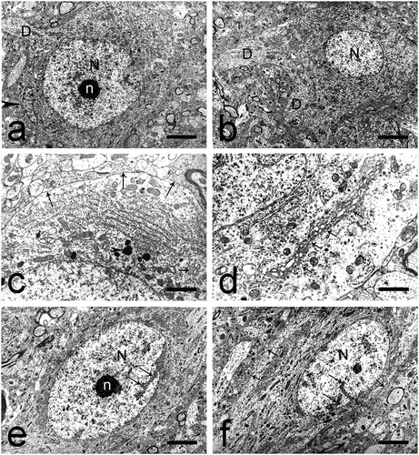
Fig. 2. Multipolar and fusiform large neurons in the dorsal claustrum in cat. (a) Multipolar large neuron with an oval nucleus (N). n – nucleolus; D – dendrite; black arrowhead – glial cell. Scale bar − 3 μm. (b) Multipolar large neuron with an oval nucleus, electron-lucent nucleus (N). D – dendrites. Scale bar − 5.5 μm. (c) Multipolar large neuron with a typical Nissl body. Long black arrows – axo-somatic synaptic contacts; short black arrow – Golgi complex; black arrowhead – lysosomes. Scale bar − 2 μm. (d) Multipolar large neuron with prominent Golgi complex comprising cisterns and small electron-lucent vesicles (black arrows). Scale bar − 1.4 μm. (e) Fusiform large neuron with an oval nucleus (N) and an electron-dense nucleolus (n) containing fibrillar centers. The nuclear membrane forms invaginations of various sizes (long black arrows). Scale bar − 5.5 μm. (f) Fusiform large neuron with a slightly elongated nucleus (N). Long black arrows – invaginations of the nuclear membrane; short black arrows – axo-somatic synaptic contacts. Scale bar − 5.5 μm.

Fig. 3. Cylindrical large neurons and large neurons of irregular shape in the dorsal claustrum in cat. (a) Cylindrical large neuron with a bean-shaped nucleus (N). The nuclear membrane forms deep invaginations (long black arrows), giving the nucleus a lobulated appearance. Scale bar − 5.5 μm. (b) Cylindrical large neuron containing a typical nucleolus-resembling structure (NucRS), recognized as a nematosome. Typical Nissl bodies are also seen. Scale bar − 1.2 μm. (c) Cylindrical large neuron containing a nematosome with an electron-dense central portion and electron-lucent peripheral portion. Long black arrows – axo-somatic synaptic contacts. Scale bar − 1.3 μm. (d) A special type of nematosome with an electron-lucent central portion built of amorphous material and a large number of filaments. Scale bar − 1.1 μm. (e) Large neuron of irregular shape. The nuclear membrane forms deep invaginations (long black arrows), giving the nucleus (N) a lobulated appearance. Black arrowhead – glial cell. Scale bar − 5.5 μm.

Fig. 3. Cylindrical large neurons and large neurons of irregular shape in the dorsal claustrum in cat. (a) Cylindrical large neuron with a bean-shaped nucleus (N). The nuclear membrane forms deep invaginations (long black arrows), giving the nucleus a lobulated appearance. Scale bar − 5.5 μm. (b) Cylindrical large neuron containing a typical nucleolus-resembling structure (NucRS), recognized as a nematosome. Typical Nissl bodies are also seen. Scale bar − 1.2 μm. (c) Cylindrical large neuron containing a nematosome with an electron-dense central portion and electron-lucent peripheral portion. Long black arrows – axo-somatic synaptic contacts. Scale bar − 1.3 μm. (d) A special type of nematosome with an electron-lucent central portion built of amorphous material and a large number of filaments. Scale bar − 1.1 μm. (e) Large neuron of irregular shape. The nuclear membrane forms deep invaginations (long black arrows), giving the nucleus (N) a lobulated appearance. Black arrowhead – glial cell. Scale bar − 5.5 μm.

Fig. 4. Medium-sized neurons in the dorsal claustrum in cat. (a) Medium-sized neuron of elliptical shape with an elongated electron-lucent nucleus (N) and a centrally located nucleolus (n) with reticular structure. Scale bar − 3 μm. (b) Cilium in a medium-sized elliptical neuron (black arrowheads). Scale bar − 1.4 μm. (c) Cilium in a medium-sized elliptical neuron (black arrowheads). Scale bar − 1.6 μm. (d) Medium-sized neuron of irregular shape with a large, electron-lucent nucleus (N). The nuclear membrane forms wide shallow invaginations (long black arrows). The nucleolus (n) is relatively large, eccentrically located, with a reticular structure. Scale bar − 4 μm.
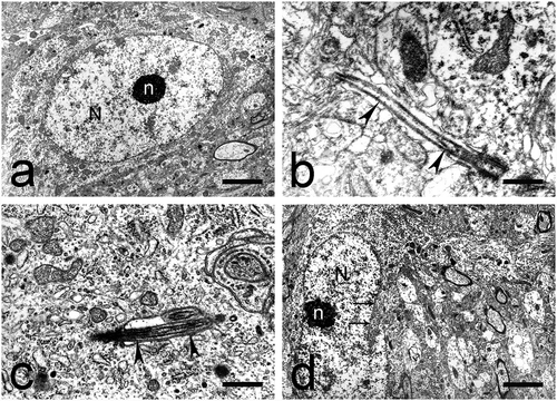
Fig. 4. Medium-sized neurons in the dorsal claustrum in cat. (a) Medium-sized neuron of elliptical shape with an elongated electron-lucent nucleus (N) and a centrally located nucleolus (n) with reticular structure. Scale bar − 3 μm. (b) Cilium in a medium-sized elliptical neuron (black arrowheads). Scale bar − 1.4 μm. (c) Cilium in a medium-sized elliptical neuron (black arrowheads). Scale bar − 1.6 μm. (d) Medium-sized neuron of irregular shape with a large, electron-lucent nucleus (N). The nuclear membrane forms wide shallow invaginations (long black arrows). The nucleolus (n) is relatively large, eccentrically located, with a reticular structure. Scale bar − 4 μm.

Fig. 5. Round and elliptical small neurons in the dorsal claustrum in cat. (a) Round small neuron with a large, electron-lucent nucleus (N). The nuclear membrane forms deep invaginations (long black arrows). The nucleolus (n) is eccentrically located, with prominent fibrillar centers. A rod-shaped intranuclear inclusion (black arrowhead) is also seen. Short black arrow – electron-dense inclusion in the cytoplasm. Scale bar − 4.5 μm. (b) Rod-shaped intranuclear inclusion (black arrowheads) built of fine granular material and parallel fine fibrils. Scale bar − 1.3 μm. (c) Rod-shaped intranuclear inclusions (black arrowheads) built of bundles of filaments. N – nucleus. Scale bar − 4.5 μm. (d) Rod-shaped intranuclear inclusion (black arrowheads) with a crystal-like structure. Scale bar − 1.3 μm. (e) Round small neuron with a large, electron-lucent nucleus (N) and a centrally located nucleolus (n). The nuclear membrane forms deep invaginations (long black arrows). Tight junctions (short black arrows) are seen between two neurons and one neuron and one glial cell (black arrowhead). Scale bar − 4.5 μm. (f) Elliptical small neuron with an oval-shaped, electron-lucent nucleus (N). The nuclear membrane forms a small number of invaginations (long black arrows). The nucleolus (n) is centrally located, has an oval shape and a compact structure. Short black arrows – tight junctions. Scale bar − 3 μm.
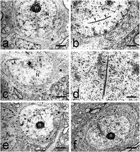
Fig. 5. Round and elliptical small neurons in the dorsal claustrum in cat. (a) Round small neuron with a large, electron-lucent nucleus (N). The nuclear membrane forms deep invaginations (long black arrows). The nucleolus (n) is eccentrically located, with prominent fibrillar centers. A rod-shaped intranuclear inclusion (black arrowhead) is also seen. Short black arrow – electron-dense inclusion in the cytoplasm. Scale bar − 4.5 μm. (b) Rod-shaped intranuclear inclusion (black arrowheads) built of fine granular material and parallel fine fibrils. Scale bar − 1.3 μm. (c) Rod-shaped intranuclear inclusions (black arrowheads) built of bundles of filaments. N – nucleus. Scale bar − 4.5 μm. (d) Rod-shaped intranuclear inclusion (black arrowheads) with a crystal-like structure. Scale bar − 1.3 μm. (e) Round small neuron with a large, electron-lucent nucleus (N) and a centrally located nucleolus (n). The nuclear membrane forms deep invaginations (long black arrows). Tight junctions (short black arrows) are seen between two neurons and one neuron and one glial cell (black arrowhead). Scale bar − 4.5 μm. (f) Elliptical small neuron with an oval-shaped, electron-lucent nucleus (N). The nuclear membrane forms a small number of invaginations (long black arrows). The nucleolus (n) is centrally located, has an oval shape and a compact structure. Short black arrows – tight junctions. Scale bar − 3 μm.

Fig. 6. Elongated small neurons and neurogliaform (‘dwarf’) cells in the dorsal claustrum in cat. (a) Elongated small neuron with a large, electron-lucent nucleus (N). The nuclear membrane forms various invaginations (long black arrow). The nucleolus (n) is centrally located with prominent nucleolus-associated chromatin. The Golgi complex is represented by small cisterns and electron-lucent vesicles (short black arrow). Black arrowhead – cilium protruding from the cellular surface. Scale bar − 4.5 μm. (b) Elongated small neuron with a large, electron-lucent nucleus (N). The nuclear membrane forms extensive invaginations which form several branches (long black arrows). The nucleolus (n) is centrally located with prominent nucleolus-associated chromatin. Scale bar − 4.5 μm. (c) Terminal boutons, forming symmetrical axo-somatic synaptic contacts (long black arrows). N – nucleus; n – nucleolus. Scale bar − 1.3 μm. (d) Two neurogliaform (‘dwarf’) cells with electron-lucent nuclei (N) and centrally located nucleoli (n) with reticular structure. Scale bar − 5.5 μm. (e) Neurogliaform (‘dwarf’) cell with an irregularly-shaped nucleus (N) owing to the numerous invaginations of the nuclear membrane (long black arrows). n – nucleolus. Scale bar − 4.5 μm. (f) Axo-somatic synaptic contact on the surface of a neurogliaform (‘dwarf’) cell (short black arrow). N – nucleus. Scale bar − 1.6 μm.
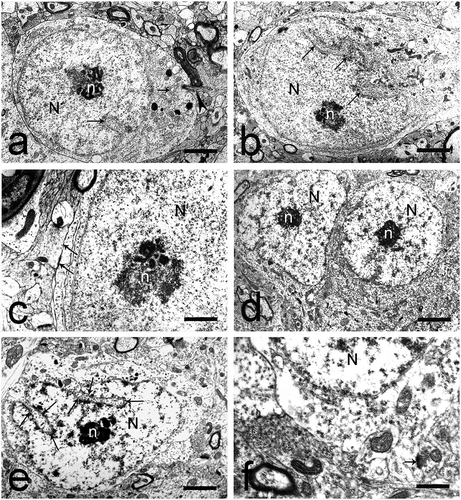
Fig. 6. Elongated small neurons and neurogliaform (‘dwarf’) cells in the dorsal claustrum in cat. (a) Elongated small neuron with a large, electron-lucent nucleus (N). The nuclear membrane forms various invaginations (long black arrow). The nucleolus (n) is centrally located with prominent nucleolus-associated chromatin. The Golgi complex is represented by small cisterns and electron-lucent vesicles (short black arrow). Black arrowhead – cilium protruding from the cellular surface. Scale bar − 4.5 μm. (b) Elongated small neuron with a large, electron-lucent nucleus (N). The nuclear membrane forms extensive invaginations which form several branches (long black arrows). The nucleolus (n) is centrally located with prominent nucleolus-associated chromatin. Scale bar − 4.5 μm. (c) Terminal boutons, forming symmetrical axo-somatic synaptic contacts (long black arrows). N – nucleus; n – nucleolus. Scale bar − 1.3 μm. (d) Two neurogliaform (‘dwarf’) cells with electron-lucent nuclei (N) and centrally located nucleoli (n) with reticular structure. Scale bar − 5.5 μm. (e) Neurogliaform (‘dwarf’) cell with an irregularly-shaped nucleus (N) owing to the numerous invaginations of the nuclear membrane (long black arrows). n – nucleolus. Scale bar − 4.5 μm. (f) Axo-somatic synaptic contact on the surface of a neurogliaform (‘dwarf’) cell (short black arrow). N – nucleus. Scale bar − 1.6 μm.