Figures & data
Figure 1: A is an H&E photomicrograph of the patient’s endometrial curettage showing high magnification of the haematolymphoid neoplasm. B shows diffuse, strong positive CD20 staining of neoplastic B-cells. C shows positive CD79a staining of neoplastic B-cells. D shows focal membranous CD10 staining of neoplastic B-cells. E shows diffuse positive staining of tumour cells on a BCL2 stain. F shows negative staining of neoplastic cells with a BCL6 stain. G shows positive MUM1 staining of neoplastic cells. H demonstrates a high proliferative index on a Ki-67 stain (original magnification A-H: x400). The panel of stains seen in figures B–H allows for a pathological diagnosis of diffuse large B-cell lymphoma to be reached. The panel of stains indicates that the DLBCL is of activated B-cell subtype, which has a more aggressive course.Citation1 Distinguishing activated B-cell subtype from germinal centre cell subtype is necessary as addition of certain therapeutic drugs has shown improvement in patients with the activated B-cell type.Citation1
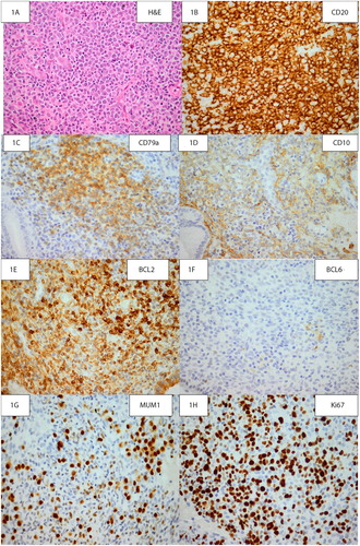
Figure 2: Electropherogram: immunoglobulin heavy chain (IgH) gene amplification by polymerase chain reaction demonstrated monoclonality with FR3 primers.