Figures & data
Table 1. Selected physico-chemical properties of soil from a potato field in Meghalaya, India
Figure 1. Dark septate endophyte, arbuscular mycorrhizal colonization and root structure of potato. (a & b) Root segments with root hairs. Bar = 1.2 mm & 1.2 mm, respectively; (c-i) portions of roots showing DSE colonization. Bar = 300μm, 100μm, 100μm, 100μm, 50μm, 50μm, & 150μm, respectively; (j-o) roots showing AMF structures. Bar = 1.2mm, 300μm, 50μm, 75μm, 50 μm & 100μm, whereas; rh-root hairs, ms-microsclerotia, dsh-dark septate hyphae, ar-arbuscules, ich-intracellular hyphae and v-vesicles.
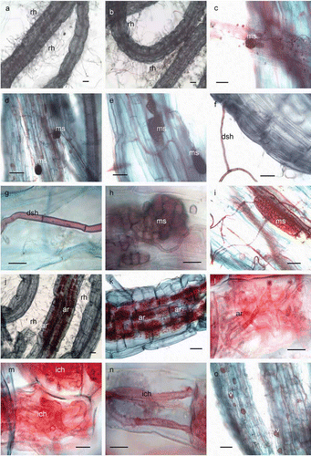
Figure 2. Arbuscular mycorrhizal and dark septate endophyte colonization in the roots of potato collected from three months.
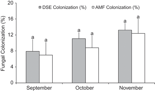
Table 2. AMF and DSE colonization of potato and monthly AMF spore density
Table 3. Correlation coefficients between fungal colonization, root hair density and soil physico-chemical properties
Figure 3. Arbuscular mycorrhizal fungi isolated from potato field. (a) Glomus macrocarpum. Bar = 200μm; (b) G. clavisporum. Bar = 150μm; (c) G. fuegianum. Bar = 200μm; (d) peridium [p] surrounding the spores of G. tortuosum. Bar = 200μm; (e) spores of G. aggregatum inside unidentified spore [us]. Bar = 500μm; (f) spores of G. microaggregatum inside cyst nematode [cs]. Bar = 500μm; (g) Glomus sp 1. Bar = 100μm; (h) Acaulospora tuberculata with sporiferous saccule [ss]. Bar = 50μm; (i) A. cavernata with sporiferous saccule. Bar = 50μm; (j) pitted ornamentation on the wall of A. cavernata. Bar = 10μm; (k) ridges on the surface of A. rehmii. Bar = 10μm; (l) wall of Pacispora chimonobambusae with clavate projections. Bar = 10μm; (m) P. bolivianawith ornamented pits. Bar = 100μm; (n) Gigaspora margarita. Bar = 400μm; (o) Scutellospora sp 2. Bar = 300μm; (p) germination shield [gs] of Scutellospora sp 2. Bar = 20μm; (q) auxiliary cells [ac] of Scutellospora sp 2. Bar = 10μm; (r) Pacispora like spore with ornamentation on the wall. Bar = 200μm; (s) hyaline layer [hl] on the wall of unidentified species. Bar = 100μm; (t) unidentified species. Bar = 100μm; (u) thorn like projection [tp] on the wall of unidentified species. Bar = 25μm; (v) wart like projection [wp] on the wall of unidentified species. Bar = 100μm & (w) hyaline layer on the wall of unidentified species. Bar = 200μm.
![Figure 3. Arbuscular mycorrhizal fungi isolated from potato field. (a) Glomus macrocarpum. Bar = 200μm; (b) G. clavisporum. Bar = 150μm; (c) G. fuegianum. Bar = 200μm; (d) peridium [p] surrounding the spores of G. tortuosum. Bar = 200μm; (e) spores of G. aggregatum inside unidentified spore [us]. Bar = 500μm; (f) spores of G. microaggregatum inside cyst nematode [cs]. Bar = 500μm; (g) Glomus sp 1. Bar = 100μm; (h) Acaulospora tuberculata with sporiferous saccule [ss]. Bar = 50μm; (i) A. cavernata with sporiferous saccule. Bar = 50μm; (j) pitted ornamentation on the wall of A. cavernata. Bar = 10μm; (k) ridges on the surface of A. rehmii. Bar = 10μm; (l) wall of Pacispora chimonobambusae with clavate projections. Bar = 10μm; (m) P. bolivianawith ornamented pits. Bar = 100μm; (n) Gigaspora margarita. Bar = 400μm; (o) Scutellospora sp 2. Bar = 300μm; (p) germination shield [gs] of Scutellospora sp 2. Bar = 20μm; (q) auxiliary cells [ac] of Scutellospora sp 2. Bar = 10μm; (r) Pacispora like spore with ornamentation on the wall. Bar = 200μm; (s) hyaline layer [hl] on the wall of unidentified species. Bar = 100μm; (t) unidentified species. Bar = 100μm; (u) thorn like projection [tp] on the wall of unidentified species. Bar = 25μm; (v) wart like projection [wp] on the wall of unidentified species. Bar = 100μm & (w) hyaline layer on the wall of unidentified species. Bar = 200μm.](/cms/asset/599883bd-bbf7-4728-bff7-d5069b14142d/tmyc_a_517787_o_f0003g.jpg)