Figures & data
Figure 1. Tumor response over time. Immunocompetent CT26 balb/c mice randomized into treatment groups, CaEP = calcium electroporation, CaUS = calcium ultrasound, BleoEP = electrochemotherapy, BleoUS = bleomycin ultrasound, Ca = calcium alone, Bleo = bleomycin alone, EP = electroporation alone, US = ultrasound alone. After one treatment tumor volume was measured over time, n = 12 (means + SD). (A) Calcium electroporated compared with control groups p < 0.001 (B) Calcium ultrasound compared with control groups p < 0.001 (C) electrochemotherapy compared with control groups p < 0.001 (D) Bleomycin ultrasound compared with control groups p < 0.001.
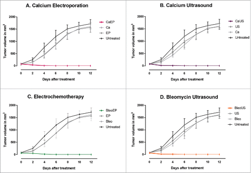
Figure 2. Survival curves after re-challenge. Surviving mice and naïve mice were re-challenged with the same tumor cell type (CT26) or a different tumor cell type (4T1) and observed for 50 d after inoculation. CT26 CaEP /4T1 CaEp = mice treated with calcium electroporation, inoculated with CT26 and 4T1, respectively; CT26 CaUS/4T1 CaUS = mice treated with calcium ultrasound, inoculated with CT26 and 4T1, respectively; CT26 BleoEP/4T1 BleoEP = mice treated with electrochemotherapy, inoculated with CT26 and 4T1, respectively; CT26 BleoUS/4T1 BleoUS = mice treated with bleomycin ultrasound, inoculated with CT26 and 4T1, respectively; CT26 naïve/4T1 naïve = naïve mice inoculated with CT26 and 4T1, respectively. N = 5 in each group. (A) Calcium electroporation compared with control groups, p = 0.004–0.008 (B) Calcium ultrasound compared with control groups, p = 0.006–0.008 (C) electrochemotherapy compared with control groups, p = 0.004–0.009 (D) Bleomycin ultrasound compared with control groups, p = 0.002–0.007.
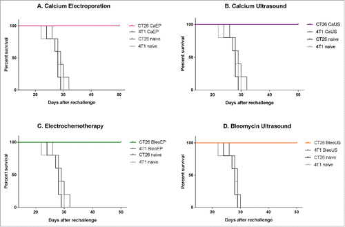
Figure 3. Heat map demonstrating the level of proinflammatory cytokines in serum 3 and 7 d after treatment. The color bar on the left represent values fold increase relative to untreated control. Values above 4-fold are represented as white. (1) Calcium electroporation showed no difference in cytokine level day 3 (p = 0.5) but a significant increased level day 7 (p<0.0001). (2) Electrochemotherapy showed a significant increase in cytokine level day 3 (p = 0.003) but no difference at day 7 (p = 1). (3) Electroporation alone showed a significant increase in cytokines both day 3 (p<0.0001) and day 7 (p<0.0001) and a noticeable increase at day 3 of G-CSF. (4) Calcium alone and bleomycin alone showed no difference in cytokine level day 3 (p = 1) but a significantly increased level day 7 (p<0.0001). (5) treatment with ultrasound showed no significant difference day 3 or 7. More detailed graph is shown in supplementary 1.
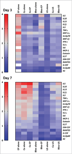
Figure 4. (A) Tumor response over time. NMRI-Foxn1nu mice randomized into treatment groups, CaEP = calcium electroporation, BleoEP = electrochemotherapy, Ca = calcium alone, Bleo = bleomycin alone, EP = electroporation alone, n = 11–12 (means + SD). After one treatment tumor volume was measured over time. The individual graphs terminates when n < 4. The difference in tumor volume between the six groups was not statistical significant (p = 0.07–1.0). (B) Survival curves from the same treatment groups show no statistical difference in survival between untreated mice and the five treatment groups (p = 0.06–0.8).
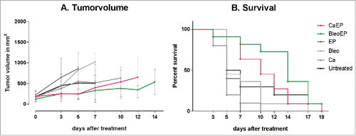
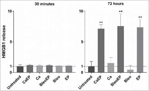
Figure 5. HMGB1 release measured in the supernatant of CT26 cells treated with CaEP = calcium electroporation, BleoEP = electrochemotherapy, Ca = calcium alone, Bleo = bleomycin alone, EP = electroporation alone, n = 4 (normalized means + SD). The supernatants was collected 30 min and 72 hafter treatment. After 30 min there no significant difference in extracellular HMGB1 concentration between the groups. At 72 h an 7-fold increase is observed for Calcium electroporation, electrochemotherapy and electroporation alone (p = 0.029) there was no statistical increase in calcium and bleomycin alone (p = 0.2).