Figures & data
Figure 1. (a) Passive targeting of nanovaccines to DCs. Nanoparticles up to 200 nm can diffuse from the interstitial fluid across the lymphatic endothelium (red) into lymph vessels. Then nanovaccines are transported to lymph nodes and targeting local DCs. Nanoparticles larger than 500 nm cannot cross the endothelium and are trapped at the injection place then skin DCs (green) can take up the nanoparticles and transport them to the lymph node for antigen presentation to T cells through dermal DCs. (b) Active targeting of nanovaccines to DCs includes nanoparticles with ligands or antibodies that bind specifically to DC surface receptors, thus directing nanovaccine uptake toward DCs. Adapted from Paulis et al. (Citation2013).
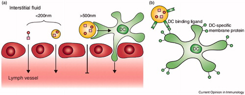
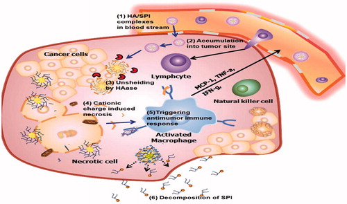
Figure 2. After the HA/SPI complexes react within the tumor tissue in response to the overexpressed HAase, the charge recovery (from negative to positive) happens. The positive charge of SPI, disrupts the integrity of the cell plasma membrane and induces necrosis. The released organelles activate the macrophages at the tumor location. The activated macrophages release cytokines that employee lymphocytes, activated natural killer cells and cytotoxic T lymphocytes. Finally, SPI decomposes into small molecule compounds for excretion without inducing systemic toxicity. Adapted from Yim et al. (Citation2014).