Figures & data
Scheme 1. Synthesis and chemical structure of P(NIPAM-co-DMA).
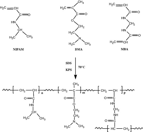
Figure 1. 1H NMR spectrum of P(NIPAM-co-DMA).
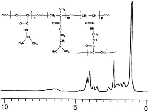
Figure 2. IR spectrum of P(NIPAM-co-DMA).
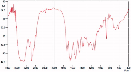
Figure 3. SEM micrographs of P(NIPAM-co-DMA) nanogel (A) and cisplatin-loaded P(NIPAM-co-DMA)/Fe3O4 nanocomposite (B).
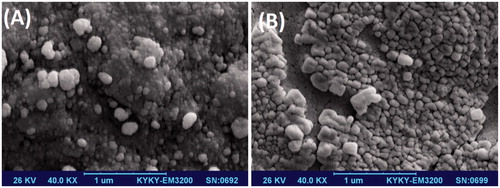
Figure 4. Size distribution of P(NIPAM-co-DMA) nanogel (A) and cisplatin-loaded P(NIPAM-co-DMA)/Fe3O4 nonocomposite (B) analysed by DLS.
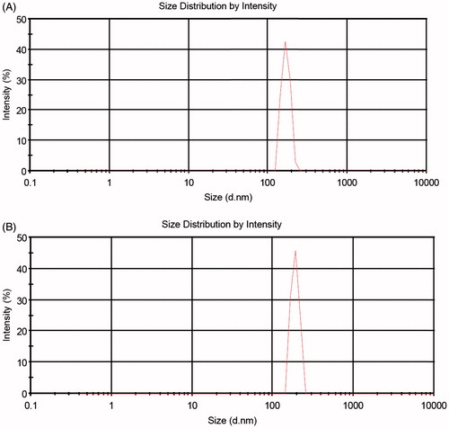
Figure 5. Magnetization curves of Fe3O4 magnetic nanoparticles (A) and cisplatin-loaded P(NIPAM-co-DMA)/Fe3O4 nanocomposite (B).
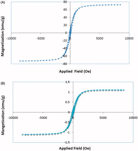
Figure 6. Separation-redispersion behaviour of cisplatin-loaded P(NIPAM-co-DMA)/Fe3O4 nanocomposite: without external magnetic field (left), with external magnetic field (right).
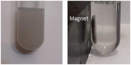
Table 1. Swelling ratio of the P(NIPAM-co-DMA) nanogel at two different temperature and pH values.
Figure 7. (A) The release behaviour of cisplatin-loaded P(NIPAM-co-DMA) nanogel. (B) The release behaviour of cisplatin-loaded P(NIPAM-co-DMA)/Fe3O4 nanocomposite.
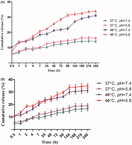
Figure 8. The HepG2 cells in cultivation medium (A). The cytocompatibility checking of P(NIPAM-co-DMA) nanogel using MTT assay which showed no significant cytotoxicity to HepG2 cells (B).
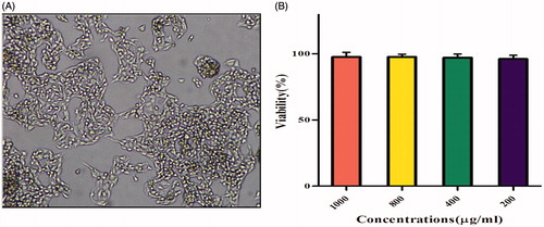
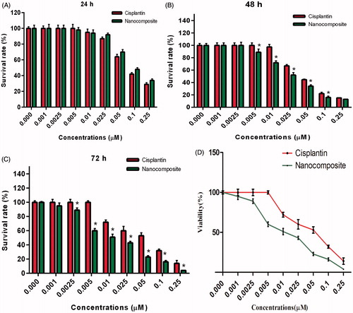
Figure 9. Growth inhibition rates by different concentration of free cisplatin and cisplatin-loaded P(NIPAM-co-DMA)/Fe3O4 nanocomposite after 24 (A), 48 (B) and 72 h (C). IC50 reduced to lower doses 72 h post incubation (D).